Warning: mkdir(): Permission denied in /home/virtual/lib/view_data.php on line 81 Warning: fopen(/home/virtual/e-kjs/journal/upload/ip_log/ip_log_2024-04.txt): failed to open stream: No such file or directory in /home/virtual/lib/view_data.php on line 83 Warning: fwrite() expects parameter 1 to be resource, boolean given in /home/virtual/lib/view_data.php on line 84 Angiolipoma on the Lumbar Spine
Korean J Spine Search

CLOSE


Kang, Kim, Kim, and Park: Angiolipoma on the Lumbar Spine

Abstract

Angiolipomas are composed of mature adipose tissue and blood vessels. Spinal angiolipomas are rare benign tumors with a good prognosis, and most symptoms are due to progressive compression of the spinal cord and root. The treatment of choice is total resection without any additional therapy. We report a case of an epidural angiolipoma on the lumbar spine in a 69-year-old man with weakness of the lower extremities. Lumbar magnetic resonance images revealed a well-enhanced epidural mass. He underwent laminectomy, and the tumor was completely removed. Pathologic examination confirmed the tumor was an angiolipoma. The patient’s symptoms gradually improved postoperatively.

INTRODUCTION

Angiolipomas are benign tumors that usually occur in the subcutaneous layer of the trunk and extremities. They rarely occur on the spine. Spinal angiolipomas commonly occur on the midthoracic spine and are located on the dorsolateral aspect of the cord3). The symptoms of spinal angiolipomas usually appear gradually because of the progressive compression of the cord. However, sometimes, symptoms can arise rapidly because of intratumoral hemorrhage and venous thrombosis. Most spinal angiolipomas have a good prognosis following surgical resection, and noninfiltrating tumors are relatively easily dissected from the dura and adjacent structures because of the good encapsulation of the mass1,10). In contrast, infiltrating tumors are entirely or partially unencapsulated12).
We report a rare case of a spinal angiolipoma on the lumbar spine, in which treatment using surgical removal resulted in a good outcome.

CASE REPORT

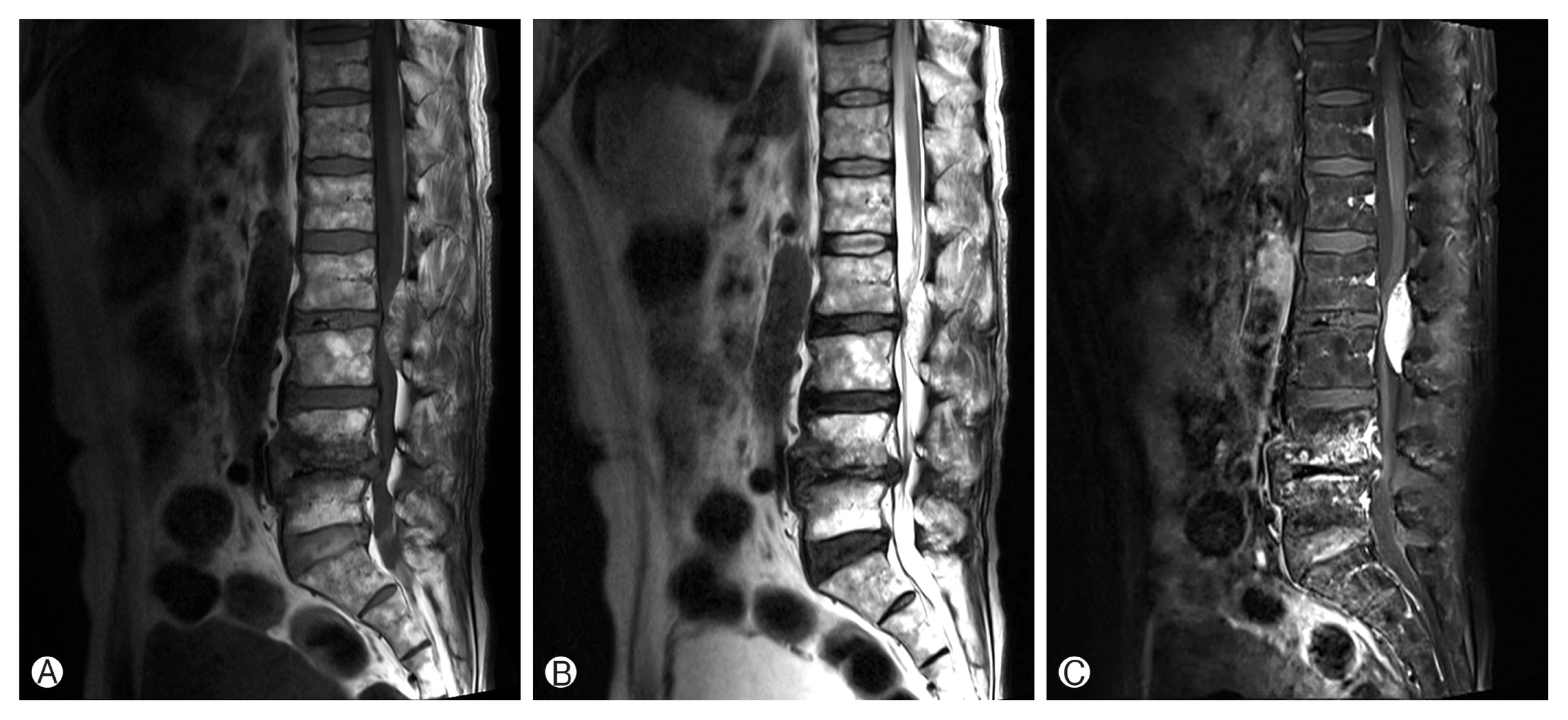
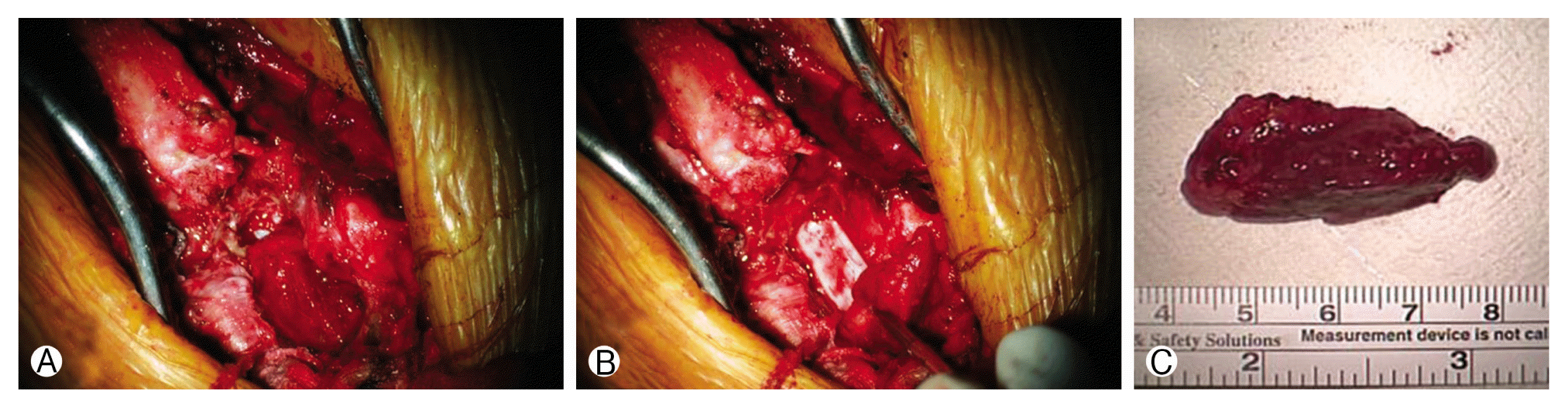
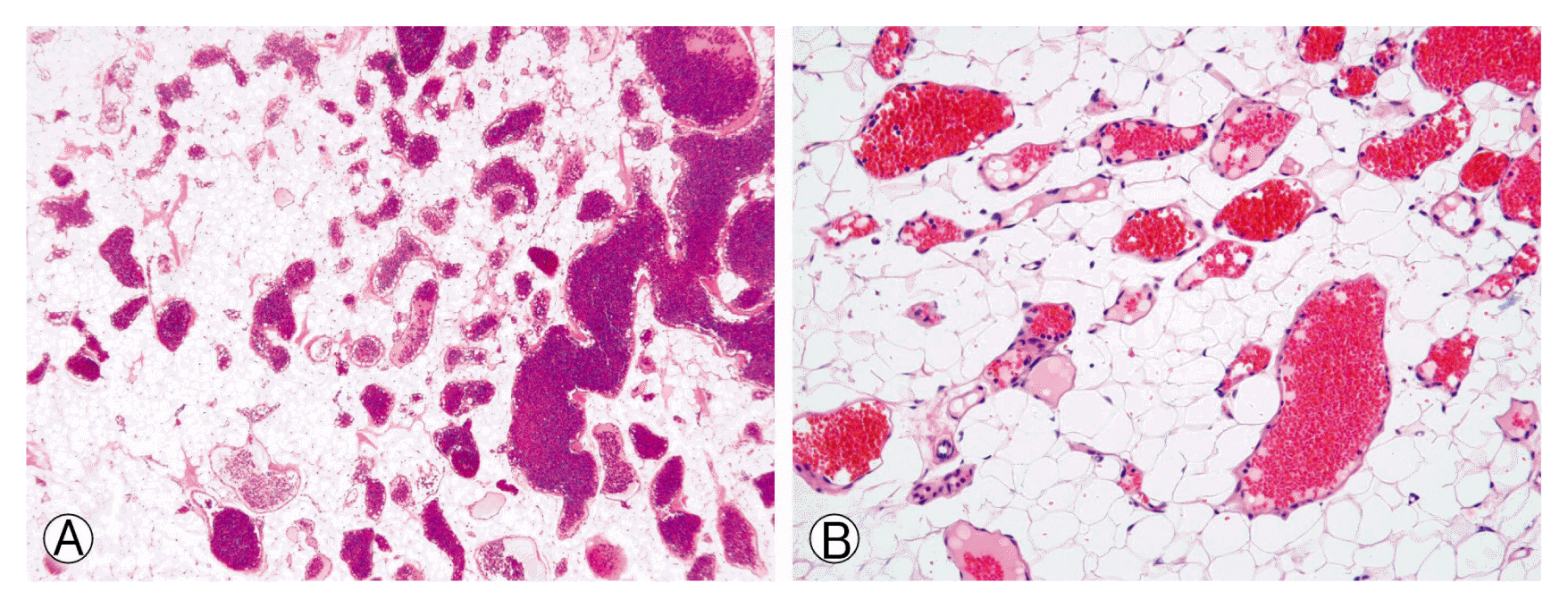
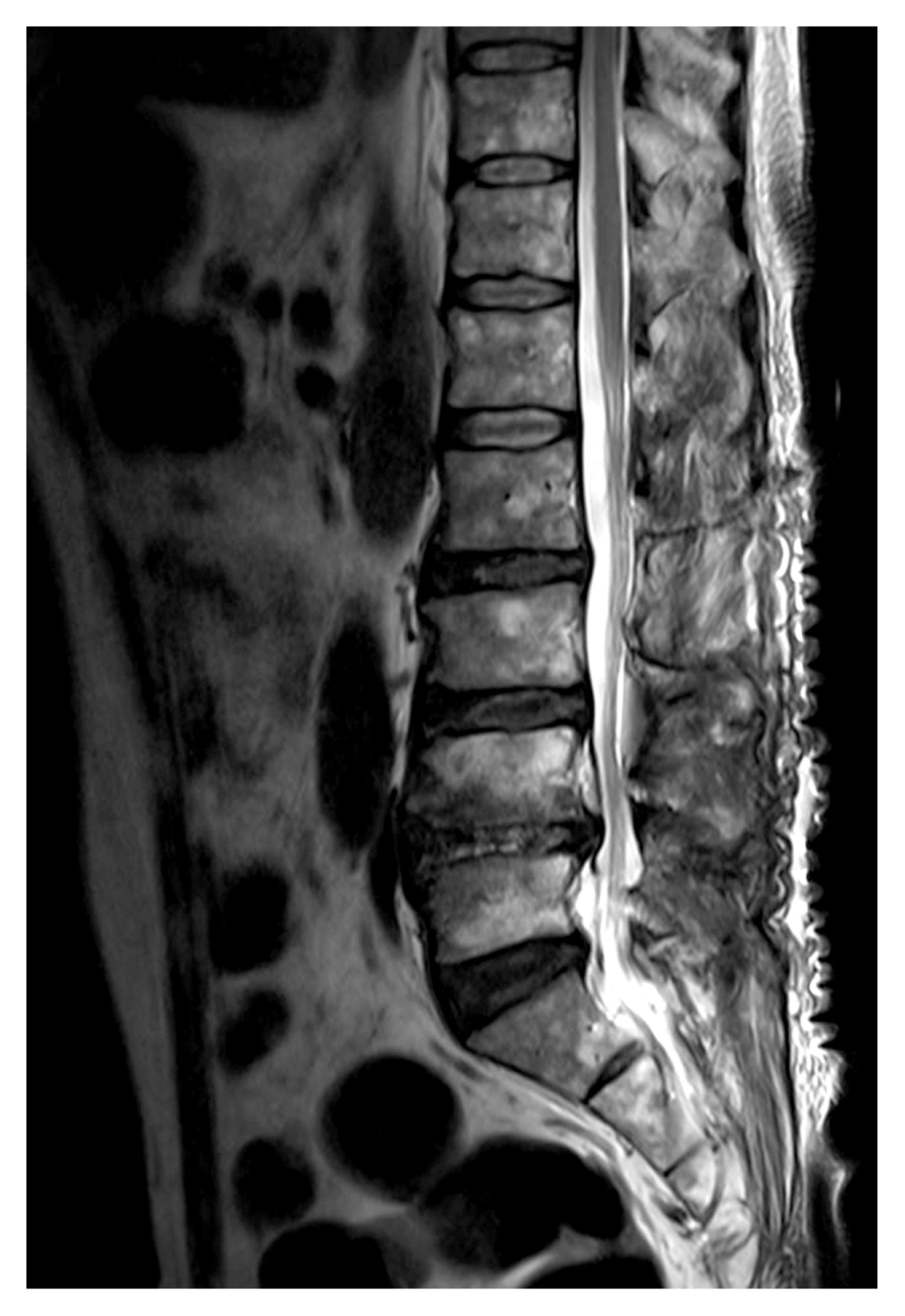
A 69-year-old man was admitted to our department with long-lasting numbness and subacute-onset weakness of both lower extremities. His weakness developed 1 week prior to presentation, and was especially evident in his hip joint. Lumbar computed tomography revealed a prominent epidural mass with dural compression at the level of L2–3 and severe canal stenosis at L4–5. Magnetic resonance imaging (MRI) revealed a well-defined mass with strong enhancement. The mass was hyperintense on T2-weighted imaging (WI) and isointense to slightly hyperintense on T1-WI (Fig. 1). We pre sumed the weakness developed because of the mass lesion. We performed a total laminectomy of L2 with removal of the mass, and used a unilateral approach for bilateral decompression at L4–5. The mass was reddish and friable and was easily dissected from the dura (Fig. 2). It was surrounded by a blood clot, suggesting the possible occurrence of intratumoral bleeding. The mass was confirmed as an angiolipoma on pathologic examination, which showed mature adipocytes and thin-walled capillary-sized vessels (Fig. 3). Follow-up MRI showed total removal of the mass (Fig. 4). After the surgery, his weakness gradually improved, and he was discharged without additional treatment. His weakness fully recovered 3 months later.

DISCUSSION

Angiolipomas are usually composed of mature lipomatous tissue and proliferating vessels and rarely occur on the spine. Most angiolipomas are slow-growing and present with gradually progressing radicular symptoms. In patients with intratumoral hemorrhage, the symptoms can include acute-onset myelopathy2). Our patient had bleeding around the mass, and the development of lower extremity weakness was relatively subacute. Increased venous pressure, obstructed venous drainage, increased adiposity, and hormonal changes may influence the size of the mass3). Pregnancy may be an aggravating factor5).
On computed tomography, angiolipomas appear hypointense relative to the spinal cord13). Most angiolipomas appear isointense or hyperintense on T1-WI and hyperintense on T2-WI, with good enhancement3,4). The same pattern was also observed in our patient. A mass appearing hypointense on T1-WI may suggest that a vascular lesion is more likely than is an adipose lesion8,14), despite the absence of signal voids. Angiolipomas composed of more lipomatous tissue (>50%) have a trabeculated or mottled appearance. However, those composed of more vascular components show large foci in the mass on magnetic resonance images8).
Angiolipomas are composed of mature adipose tissue and blood vessels and are differentiated from capillary angiomas and cavernous angiomas3). Immunohistochemical studies on angiolipomas show positive findings for CD34 and smooth muscle actin7,15).
Angiolipomas are classified as either noninfiltrating or infiltrating. Noninfiltrating angiolipomas are more common6,10), and are relatively easily dissected from the dura. However, if MRI suggests a highly vascular lesion, preoperative angiography and embolization can be performed9). Noninfiltrating tumors are usually located in the posterior epidural space, and infiltrating tumors are generally located in the anterior epidural space4). Infiltrating angiolipomas can also invade the vertebral body and paraspinal area7). Sometimes, infiltrating angiolipomas may be mistaken for malignant tumors because of the invasion of surrounding tissue10,16).
Total surgical resection is the treatment of choice, but total removal of the mass is difficult in cases of infiltrating angiolipomas. Noninfiltrating angiolipomas have a good prognosis after total surgical resection, and infiltrating angiolipomas have a good prognosis after incomplete resection6,12). Previously, wider resection including the surrounding tissue was recommended for infiltrating angiolipomas11). However, there is no difference in the outcomes between both types of angiolipomas6,15). Recurrence is rare after surgical resection, even in cases of subtotal resection3) and absence of malignant transformation13).

CONCLUSION

We report a rare case of noninfiltrating spinal angiolipoma, in which treatment using total surgical resection resulted in a good outcome. Follow-up evaluation is required to identify rare cases of recurrence.

CONFLICT OF INTEREST

CONFLICT OF INTEREST

No potential conflict of interest relevant to this article was reported.

Fig. 1
Sagittal lumbar magnetic resonance images showing the epidural mass at the L2–3 level. (A) Isointense to slightly hyperintense lesion on the T1-weighted image (WI), (B) hyperintense lesion on the T2-WI, and (C) good enhancement after gadolinium administration.
kjs-14-3-112f1.gif
Fig. 2
The photographs show a 4×1.5-cm-sized reddish, friable epidural mass (A, C), which is easily dissected from the dura (B).
kjs-14-3-112f2.gif
Fig. 3
Histological examination reveals mature adipocytes and thin-walled capillary-sized vessels (H&E staining: A, ×40; B, ×200).
kjs-14-3-112f3.gif
Fig. 4
Postoperative lumbar magnetic resonance image showing total resection of the tumor.
kjs-14-3-112f4.gif

REFERENCES

1. Choi KC, Jang KS, Chough CK, Rha HK. Lumbosacral extradural angiolipoma: a case report. Korean J Spine 2:157-159. 2005.

2. da Costa MD, de Paz DA, Rodrigues TP, Gandolfi AC, Lamis FC, Stavale JN, et al. Hemorrhagic onset of spinal angiolipoma. J Neurosurg Spine 21:913-915. 2014.
crossref pmid
3. Gelabert-González M, García-Allut A. Spinal extradural angiolipoma: report of two cases and review of the literature. Eur Spine J 18:324-335. 2009.
crossref pmid pmc
4. Ghanta RK, Koti K, Dandamudi S. Spinal epidural angiolipoma: a rare cause of spinal cord compression. J Neurosci Rural Pract 3:341-343. 2012.
crossref pmid pmc
5. Glynn D, Murray B, Cryan J, O’Brien D, Kavanagh E. Spinal epidural angiolipoma. Spine J 16:e531-532. 2016.
crossref pmid
6. Guzey FK, Bas NS, Ozkan N, Karabulut C, Bas SC, Turgut H. Lumbar extradural infiltrating angiolipoma: a case report and review of 17 previously reported cases with infiltrating spinal angiolipomas. Spine J 7:739-744. 2007.
crossref pmid
7. Han SR, Yee GT, Choi CY, Lee CH. Infiltrating spinal angiolipoma. J Korean Neurosurg Soc 52:161-163. 2012.
crossref pmid pmc
8. Hu S, Hu CH, Hu XY, Wang XM, Dai H, Fang XM, et al. MRI features of spinal epidural angiolipomas. Korean J Radiol 14:810-817. 2013.
crossref pmid pmc
9. Konya D, Ozgen S, Kurtkaya O, Pamir NM. Lumbar spinal angiolipoma: case report and review of the literature. Eur Spine J 15:1025-1028. 2006.
crossref pmid
10. Leu NH, Chen CY, Shy CG, Lu CY, Wu CS, Chen DC, et al. MR imaging of an infiltrating spinal epidural angiolipoma. AJNR Am J Neuroradiol 24:1008-1011. 2003.
pmid pmc
11. Lin JJ, Lin F. Two entities in angiolipoma. A study of 459 cases of lipoma with review of literature on infiltrating angiolipoma. Cancer 34:720-727. 1974.
crossref pmid
12. Nadi MM, Nadi AM, Zabara MY, Ahmad TM. Management of infiltrating spinal epidural angiolipoma. Neurosciences (Riyadh) 20:159-163. 2015.
crossref pmid pmc
13. Nanassis K, Tsitsopoulos P, Marinopoulos D, Mintelis A, Tsitsopoulos P. Lumbar spinal epidural angiolipoma. J Clin Neurosci 15:460-463. 2008.
crossref pmid
14. Provenzale JM, McLendon RE. Spinal angiolipomas: MR features. AJNR Am J Neuroradiol 17:713-739. 1996.
pmid pmc
15. Si Y, Wang Z, Pan Y, Lin G, Yu T. Spinal angiolipoma: etiology, imaging findings, classification, treatment, and prognosis. Eur Spine J 23:417-425. 2014.
crossref pmid
16. Sim K, Tsui A, Paldor I, Kaye AH, Gaillard F. Four cases of spinal epidural angiolipoma. J Clin Neurosci 25:134-139. 2016.
crossref pmid


Editorial Office
CHA University, CHA School of Medicine Bundang Medical Center
59 Yatap-ro, Bundang-gu, Seongnam-si, Gyeonggi-do 13496, Korea
Tel: +82-31-780-1924  Fax: +82-31-780-5269  E-mail: support@e-neurospine.org
The Korean Spinal Neurosurgery Society
#407, Dong-A Villate 2nd Town, 350 Seocho-daero, Seocho-gu, Seoul 06631, Korea
Tel: +82-2-585-5455  Fax: +82-2-2-523-6812  E-mail: ksns1987@gmail.com
Business License No.: 209-82-62443

Copyright © The Korean Spinal Neurosurgery Society.

Developed in M2PI

Close layer
prev next