INTRODUCTION
Most spinal dumbbell tumors are neurogenic tumors, including slow-growing tumors such as ependymomas, vertebral chordomas, meningiomas, and the rare types of nerve sheath tumors, ganglioneuromas and neurofibrosarcomas3). However, in this study we report a case of solitary osteochondroma presenting itself as a dumbbell tumor compressing the cervical spinal cord and the required surgical strategy. In regard to the surgical strategy, there was some controversy over the removal of the cervical dumbbell tumor6). In this young female, we successfully performed a combined anterior oblique and posterior approach with laminoplasty.
CASE REPORT
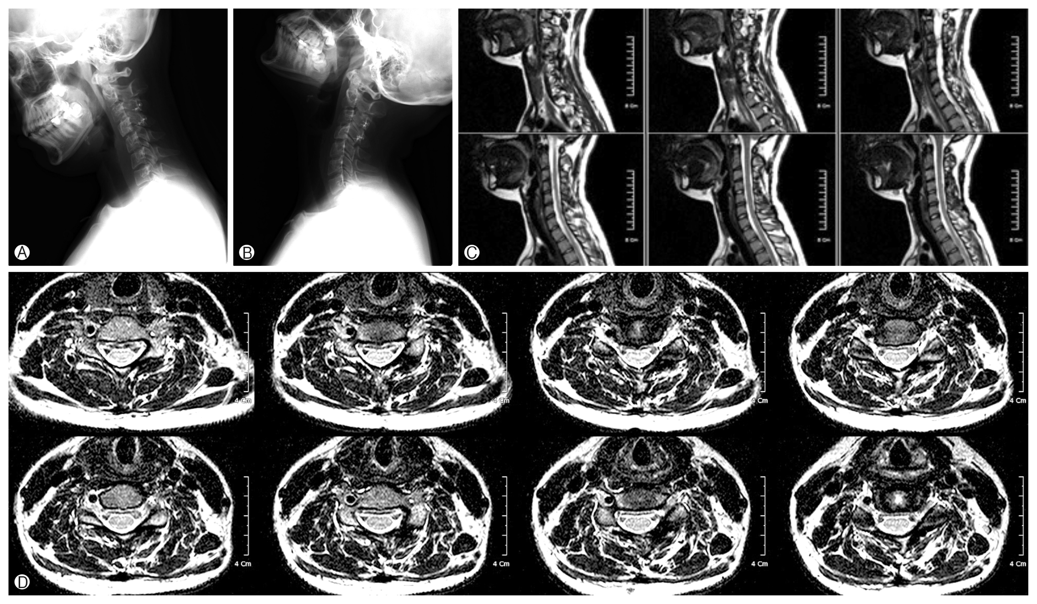
This case report was confirmed by the patient and the patient signed the informed consent form, A 16-year-old female visited our clinic complaining about progressive neck pain and left arm abduction weakness (grade III) over the past 2 years. She was examined by plain X-ray, three-dimensional (3D)-computed tomography, magnetic resonance imaging, and vertebral artery angiogram. The analyses indicated a calcified extradural mass with a size of about 3 cm├Ś2.4 cm, ill-defined, compressing the cord in the C3ŌĆō4 portion, and extending into the neural foramen with eroded vertebral body. A vertebral angiography showed a left side hypoplastic vertebral artery (Fig. 1).
There was no evidence of family history and long bone deformities revealing the multiple hereditary exostosis.
The tumor was successfully excised using a modified combined anterior oblique and posterior approach. In this approach, the extraforaminal portion of the tumor was first removed through the anterior oblique approach. For this, the patientŌĆÖs body including the neck was tilted to the right side for a proper surgical view to avoid vertebral artery injury using an intraoperative doppler. In the second part, the intracanal portion of the tumor was removed via a posterior approach with C3/C4 laminoplasty under intraoperative neuro-monitoring (Fig. 2). The tumor showed a dark-red colored leather-like consistency with some calcification.
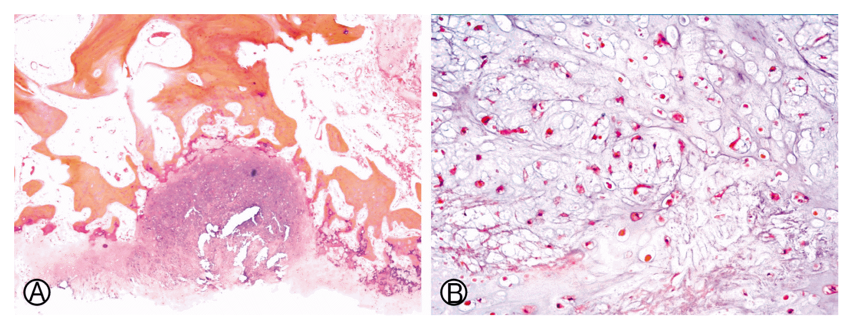
A histopathologic study of the resected material showed a cartilaginous cap and an underlying bone with enchondral ossification. The cartilaginous cap demonstrated chondrocytes without cytologic atypia comparable with osteochondroma (Fig. 3).
Postoperative, the symptoms of the female were improved. Clinical and radiological assessments were performed in the 5 years after the operation. Clinically, she was symptom-free and there was neither a spinal instability nor kyphosis or tumor recurrence in the imaging studies (Fig. 4).
DISCUSSION
Osteochondromas are common benign long bone tumors in young adults. However, they rarely involve the spinal column. Moreover, its presentation as compressive myelopathy is rare. Most of the literature of these tumors is in form of case reports.
The term ŌĆ£dumbbell tumorŌĆØ describes spinal tumors that acquire an hourglass shape in the course of their growth as they encounter an anatomic barrier such as the dura mater, a nerve root foramen, or bony elements. Various tumoral, vascular, and developmental causes may lead to dumbbell lesions5,15,17,18).
However, to our knowledge, a solitary osteochondroma presenting itself as a dumbbell tumor in the cervical spine is not reported yet. In this patient, a slow growing osteochondroma may have passed through the corresponding neural foramen, widened, and led to the so-called ŌĆ£dumbbellŌĆØ lesion.
The finding in this patient should be considered as an osteochondroma that can be included as a rare condition in the differential diagnosis of spinal dumbbell tumors16).
Osteochondromas are thought to arise via a process of progressive enchondral ossification of aberrant cartilage of a growth plate as a result of a congenital defect or trauma. In contrast, hereditary multiple exostoses may occur sporadically as a part of an autosomal dominant gene with variable expression12). In cases of the osteochondroma of the spine, the most common location is cervical4,10) and arises from the tip of a spinous or transverse process and rarely causes neurological symptoms14). Radiation-induced osteochomdroma may possibly be caused by a failure of the reserve cell layer in the epiphyseal growth zone.
According to Albrecht et al.2), 30% of spinal solitary osteochondroma lead to spinal cord compression. However, a neurologic disease is thought to be the result of progressive encroachment of the slowly expanding osteochondroma on neural structures. A similar case as ours was reported in the literature of a 65-year-old patient with a manifested dumbbell mass passing through the thoracic neural foramen leading to cord compression. The patient was treated using hemilaminectomy with subtotal tumor excision. A clinical follow-up 2 years later revealed normal findings in that case16). In our case, postoperative follow-up for the next 5 years revealed neither radiologically tumor recur nor spinal instability.
The surgical removal is mandatory if compressing myelopathy exists. Generally, a surgical outcome is good. However, a recurrence after surgery is possible in case of incomplete removal of the cartilaginous cap19). In case of a recurrence, one should anticipate the possibility of malignant transformation of the osteochondroma or of a low-grade chondrosarcoma initially poorly classified. The risk of malignant transformation of multiple osteocartilaginous exostoses is well known. However, a malignant tumor developing from a solitary osteochondroma is rarely observed2).
Current results in surgical approaches for spinal cord compression due to spinal solitary osteochondroma are generally good. For example, only 15% of patients failed to improve after surgery or even worsened by a decompressive procedure8). But there has been some controversy in regard to surgical methods of the cervical dumbbell type tumor.
Several authors recommended an anterolateral approach. The advantage of this approach is good surgical views, but it is unfamiliar to most neurosurgeons7,9). On the other hand, Lot and George13) insisted on complete excision using a lateral approach and vertebral artery control. However, a lateral approach still carries the risk of injuring not only the vertebral artery but also the phrenic, vagus, accessory, or hypoglossal nerves.
Eun6) reported a modified posterior midline exposure with laminectomy and complete facetectomy in most cases of cervical dumbbell Schwannomas. Advantages of a combined posterior and anterior approach have been described by several authors1,11).
Asazuma et al.3) recommended that imaging-based 3D characterization of the shape and location of cervical dumbbell tumors are essential for planning optimal surgery.
We attained preoperative 3D images and performed a modified combined anterior oblique approach for an eccentrical-located extraforaminal tumor excision on the left side. The body and neck of the patient were tilted to the contralateral side of the tumor. That is nearly the same position as the oblique view of the plain X-ray. It is straight vertical to the longitudinal axis of the extraforaminal part of the tumor in three dimensions. This position provided an effective surgical corridor and the shortest distance to the tumor (Fig. 2). Next, a posterior approach for the intracanal part of tumor removal was performed with laminoplasty without solid fusion. Intraoperative neuro-monitoring was performed during the operation. Although we removed the mass around the vertebral artery using the doppler, there may remain a little tumor around the vertebral artery. However, we observed neither a tumor growth nor sarcomatous change in the next 5 years after surgery. Thus, we assume that the tumor around the vertebral artery may regress while the patient is growing up. This combined approach resulted in a nearly total tumor resection without vertebral artery injury, nor instability or kyphotic change (Fig. 4).
There was a radiologic similar case of cervical neural foramen widening chondrosarcoma in the literature20). Contrary to that article, our histopathologic diagnosis was more benign nature osteochodroma that has not recurred, nor sarcomatous change during 5 years after surgery. We proposed surgical staged strategy for safe and complete removal of the tumor.
CONCLUSION
Osteochondroma arises from enchondral bone, but it rarely involves the spine. In this patient, the tumor may have arisen from the neural arch and extended into the extradural and extraforaminal space over a long period. Appearance of dumbbellshaped tumor in the spine is usually a neurogenic tumor. However, we report a solitary osteochondroma presenting itself as a cervical dumbbell tumor. In this case, we propose a modified combined anterior oblique and posterior approach with laminoplasty as an option for tumor extension. However, further observation is essential because of the possibility of recurrence and sarcomatous change.