Aortic Injury during Transforaminal Lumbar Interbody Fusion
Article information
Abstract
Aortic injury during transforaminal lumbar interbody fusion (TLIF) is a rare but severe complication. We experienced aortic injury during TLIF at L3–4 with a 59-year-old woman diagnosed with an adjacent segment disease at L3–4. Severe bleeding occurred during disc space expansion, and the blood pressure dropped to 60/40 mmHg. The patient’s vital sign stabilized after compression with gauze and Gelfoam in addition to blood transfusion. The patient was treated with endovascular repair using a percutaneous technique after intertransverse fusion at L3–4 was completed. She recovered and is being followed-up in the outpatient department.
INTRODUCTION
Transforaminal lumbar interbody fusion (TLIF) is one of the major surgical procedures for treating degenerative spinal diseases that result in instability12), and is commonly performed due to its safety and positive prognosis. Some complications of TLIF are pseudoarthrosis with loosening of the implants, dural leakage, nerve root damage, and deep infection8).
Aortic injury is another, more serious complication of TLIF. Surgeons should be cautious of aortic injury as, though infrequent, it is potentially fatal. Here, we present the case of a patient who experienced aortic injury during TLIF.
CASE REPORT
A 59-year-old woman visited the Emergency Department with a chief complaint of radiating pain from her back to her left thigh. The findings were positive in the straight leg raise test at 50° on the left side and she had motor weakness on her left lower extremity classified as grade 3 in the manual muscle test.
She had been taking medications for hypertension, diabetes mellitus, and rheumatoid arthritis including steroids for 2 years. She had undergone 2 surgeries, specifically, aneurysm neck clipping of the right anterior communicating artery 10 years earlier, and TLIF at L4–5–S1 for spondylolisthesis at L4–5–S1.
T2-weighted magnetic resonance images showed high signal intensity at the L3–4 intervertebral disc, compression of the thecal sac by an L3–4 herniated nucleus pulposus, and unclear anterior longitudinal ligaments (ALLs) (Fig. 1). We diagnosed the patient with adjacent segment disease at L3–4 and planned to perform TLIF at L3–4.
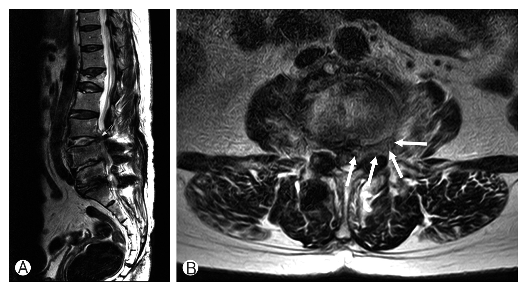
Preoperative T2-weighted magnetic resonance image of the lumbar spine. (A) The L3–4 intervertebral disc has high signal intensity on the sagittal image. (B) L3–4 disc herniation (arrows) showing protrusion to the left L3–4 foramen is indicated on the axial image.
Within the operative field, the disc had been softened by necrosis. Severe bleeding occurred during disc space expansion with a reamer for interbody fusion. We tried to control the bleeding by immediate gauze-packing. The patient’s blood pressure dropped to 60/40 mmHg and norepinephrine was administered. Blood transfusion with 3 units of packed red blood cells was performed, followed by 2 units of fresh frozen plasma to maintain stable vital signs. After bleeding control succeeded following thirty minutes of compression and Cutanplast standard (Mascia Brunelli, Milano, Italy) packing, we confirmed that her vital sign was stable and there was no bleeding or oozing around posterior longitudinal ligament (PLL) within the operative field. Then, we continued with the spinal surgery by performing intertransverse fusion instead of interbody fusion, with close monitoring of her vital signs, which remained stable.
After the spinal surgery, we performed abdominal computed tomography angiography and found a pseudoaneurysm at the L3–4 level, just 1 cm above the bifurcation of the abdominal aorta, with a size of approximately 2 cm on the right side (Figs. 2, 3).
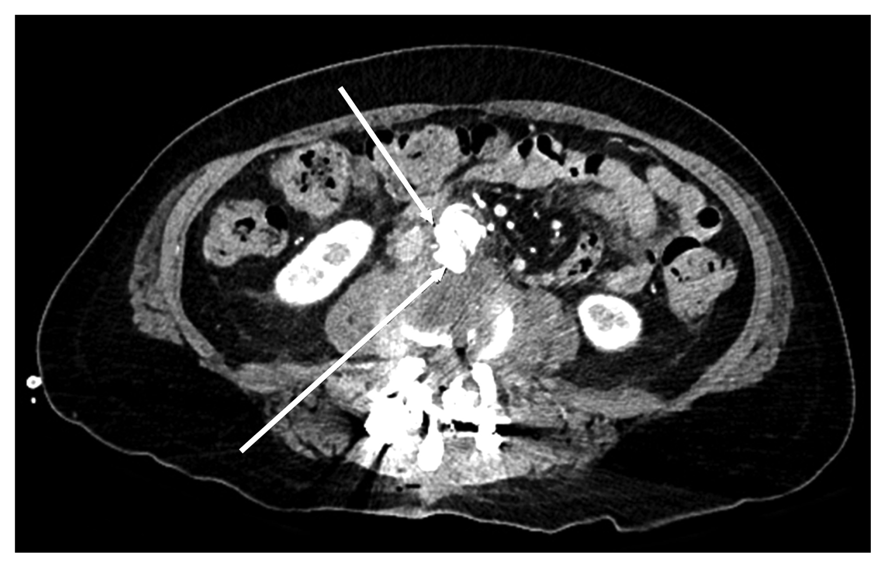
Postoperative abdominal computed tomography angiography image shows irregular lobulated contrast media collection (arrows), 2.4 cm×1.4 cm at the right side of the abdominal aorta, indicating pseudoaneurysm at the L3–4 level.
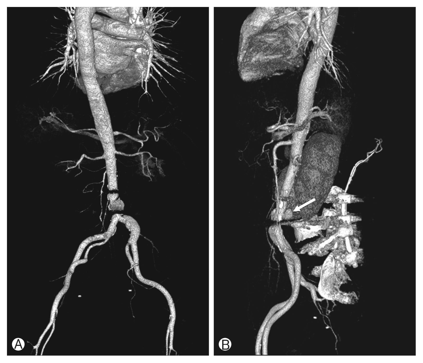
Three-dimensional reconstruction image of postoperative abdominal computed tomography angiography. The posterior-anterior view (A) and the left lateral view (B) of the abdominal aorta show pseudoaneurysm (arrow).
On the day following the spinal surgery, through consultation with a cardiothoracic surgeon and an interventional radiologist, endovascular repair was performed using a percuta neous technique, and a seal stent graft Limb-18–60 (S&G Biotech Inc., Seoul, Korea) was inserted. After obtaining angiographic confirmation of the absence of endoleakage (Fig. 4), the patient recovered well and has been visiting the outpatient department for follow-up.
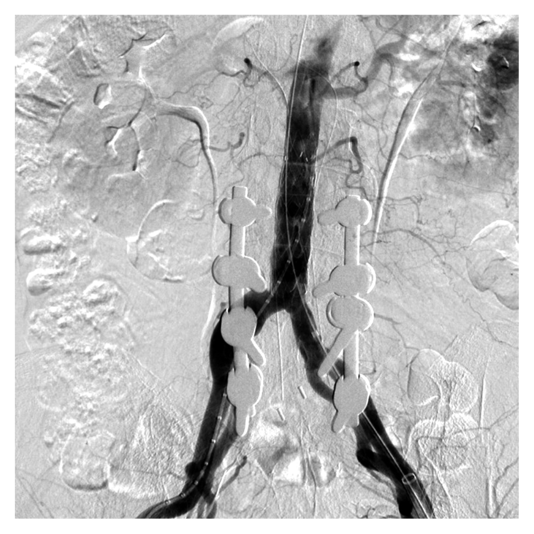
Postendovascular repair angiography shows the absence of endoleakage.
DISCUSSION
Aortic injury during TLIF is known to be a rare complication. The incidence is reported to range from 0.01%–0.06%5,10). However, it is probably more frequent because there may be many misdiagnosed cases due to the variety of clinical symptoms, making diagnosis difficult2). The mortality rate for diagnosed cases is between 15% and 65%5–7). Mortality depends on the diameter of the injured vessel, size of the tear, rapidity of diagnosis, laparotomy, and amount of control over bleeding5,7,10).
The complications of aortic injury during TLIF are divided into early and late complications. Early complications include shock due to a rupture of the major retroperitoneal vessel, while pseudoaneurysms and arteriovenous fistulas can occur years after the surgery4,9). Since anterior annulus fibrosus and ALL are tough and elastic, they can cause bleeding into the retroperitoneal space, making the bleeding difficult to see13).
Preoperative preparations—such as history taking and imaging study, including MRI —are important for prevention. Some findings should be confirmed with MRI if there is any defect on the anterior annulus fibrosus or ALL, peridiscal fibrosis with vessel adhesion and scarring following a previous abdominal surgery or radiotherapy, or pathological weakness of the vascular wall due to degeneration or erosion of the ventral disc herniation or osteophytic spurs. We suspect that there were adhesions and fibrosis within the aorta and disc caused by necrosis of the disc, and this was the reason for the aortic injury during disc expansion.
The aorta and the pelvic vessels may not have been in their normal anatomic locations or may have been tortuous due to anomalies within them. It is also recommended to check the depth and contour of the disc space, as well as any anatomic variants that could increase risk. Since the distance from the aorta to the vertebrae varies with abdominal pressure, which is affected by the position of a patient, the patient should be positioned carefully. Because the sagittal diameter of the three lowest lumbar disc ranges from 33 to 56 mm1), advancing instruments less than 3 cm from the posterior disc border may be a good method to prevent aortic injury13).
If complications occur despite efforts to prevent injury, the clinical manifestations would be abdominal pain, dyspnea, back pain, or shock3). However, it is difficult to detect these symptoms because the patient assumes a prone position under general anesthesia.
Angiography is more helpful than computed tomography scans for diagnosing aortic injury because it provides much more information about vascular anatomy and injury. Early diagnosis in addition to immediate surgical repair is necessary for this condition. While surgery has a higher mortality rate due to hemodynamic instability, endovascular surgery has better outcomes with regard to mortality and morbidity because of its minimal invasiveness3).
When aortic injury occurs, the planned spinal surgery may be postponed until the injury is completely repaired. Aortic injury during TLIF should be treated promptly11) because the injured vessel walls become weaker, making it difficult to suture the injured aorta. In addition, disseminated intravascular coagulation can occur over time.
In this case, we debated whether to complete the spinal surgery before aortic repair or to perform an aortic repair immediately. If we had stopped the spinal surgery and performed an aortic repair immediately, she would have had to undergo additional surgery for TLIF later. The patient had a sensitive and anxious personality. She was worried about having a reoperation at the adjacent segment that had previous undergone surgery on the lumbar lesion. At that moment, it was urgent to treat the aortic repair. However, considering the patient’s sensitive condition, we considered that the spinal surgery could be completed if her vital sign was stable during the surgery. Fortunately, we could control bleeding over the operative field and her vital sign kept being stable. Also, there was no oozing over PLL part and the surgery was completed. It might not be a good decision to treat the aortic injury on the next day after the spinal surgery. Because it could be dangerous if the complications of aortic injury occurred, it should be treated on the day of spinal surgery.
CONCLUSION
We present the case of a patient who experienced aortic injury during TLIF and underwent the spinal surgery before the aortic injury repair. We discovered that it is possible to complete spinal surgery if the patient is in a hemodynamically stable condition even after aortic injury.
Notes
CONFLICT OF INTEREST
No potential conflict of interest relevant to this article was reported.