Abstract
Chondromas are benign tumor of cartilaginous tissue that is rarely found in spine. The authors document a rare case of a 72 year old male patient with a cervical spinal chondroma compressing the spinal cord. The patient had symptoms of motor and sensory deficits, dysphagia and dysarthria. C1 and C2 laminotomy was done and the spinal tumor was removed. The patient gradually recovered from his previous symptom after the surgery.
-
Keywords: Spine, Neoplasm, Chondroma, Cervical spinal cord
INTRODUCTION
Chondromas are benign tumor of the cartilaginous tissue
7,
19). Spinal chondroma is very rare, especially in the cervical area
5). We report a rare case of a 72 year old male patient with cervical spine chondroma compressing spinal cord.
CASE REPORT
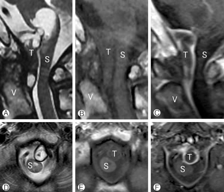
A 72 year old male patient visited the emergency room (ER) with progressive weakness and paresthesia in all extremities that were aggravated several days ago. On physical examination, the patient's motor power was decreased, showing grade III motor weakness on the right side and grade IV- on the left side. He also complained of dysarthria and dysphagia that began 12 hours before the visit to the ER. Cervical spine magnetic resonance (MR) image was taken at the ER and it showed a 3.4×1.3 cm sized heterogeneously enhancing mass with peripheral rim enhancement. The spinal tumor was based on dura and contained a cyst like portion. It was compressing the spinal cord and the exophytic cystic portion of the tumor was compressing the cervicomedullary junction (
Fig. 1).
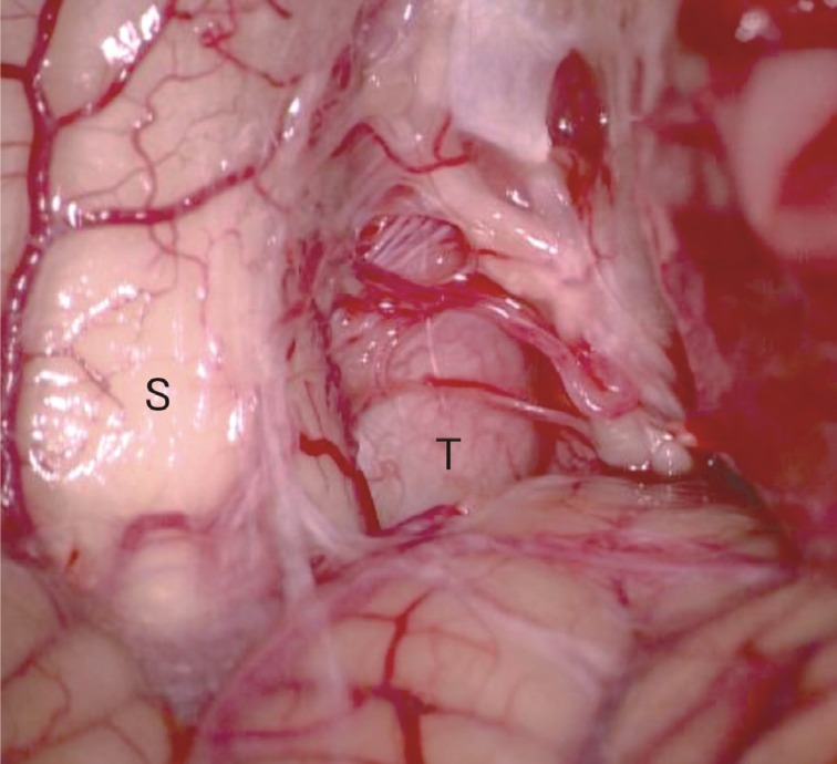
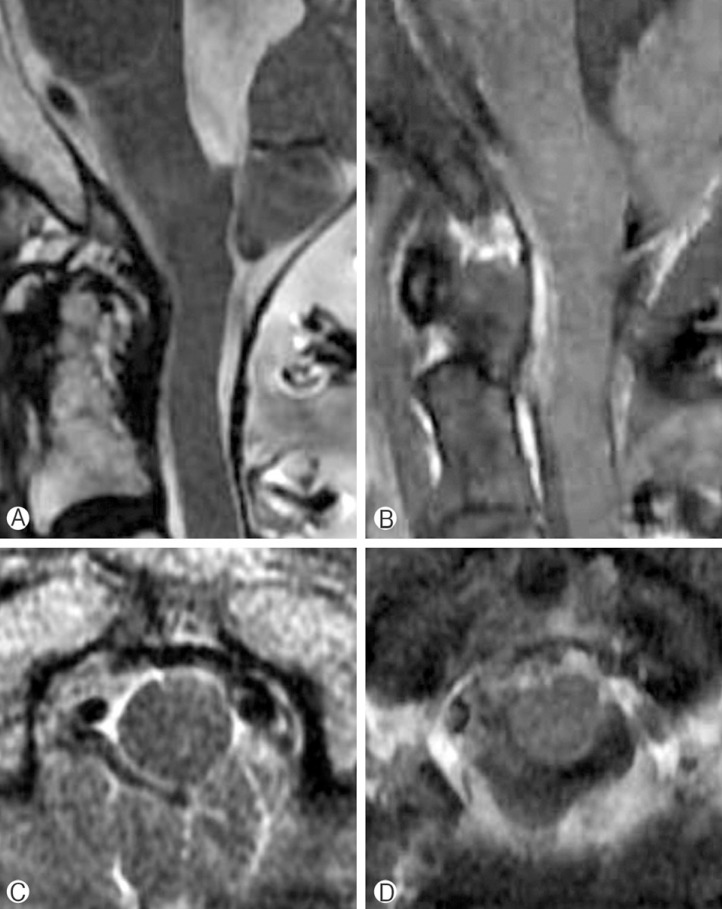
C1, C2 laminotomy was done using a conventional posterior approach. After the dura was opened, a greyish fibrous tumor compressing the spinal cord was seen outside the arachnoid membrane. The tumor was hard and was firmly attached to the ventral dura. There was no adhesion between the tumor and the spinal cord. The spinal cord was mildly deviated to the side due the ventrolaterally compressing tumor (
Fig. 2). The tumor was intralesionally excised while saving the nerve roots. Postoperative MRI showed the tumor was removed well and the spinal cord was adequately decompressed (
Fig. 3). This conventional posterior approach technique was similar in our previous report for ventral foramen magnum meningioma
16).
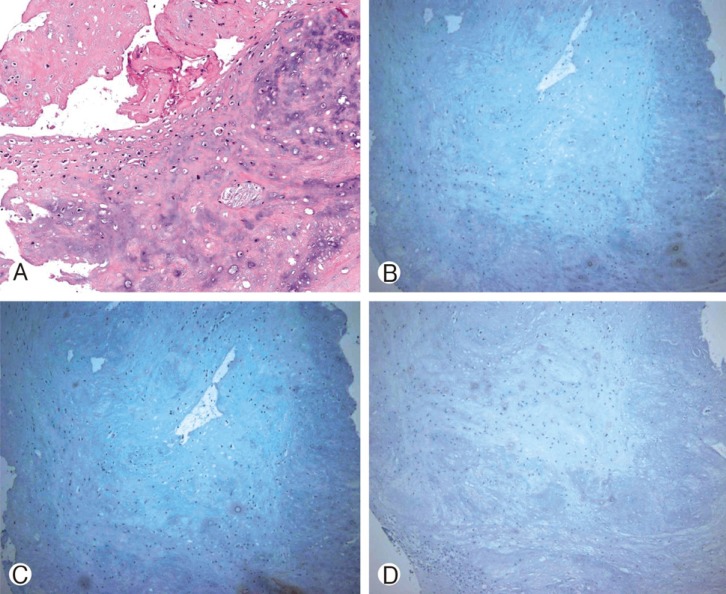
Patient's motor weakness and paresthesia was gradually improved after the surgery. The patient was able to walk using a walker three months after the surgery. Sign of dysphagia also improved over time, and he was able to swallow food without aspiration one year after surgery. The final pathology report confirmed the spinal tumor as a chondroma. Immunohistochemistry staining for cytokeratin, epithelial membrane antigen (EMA), and S-100 protein stain were all negative (
Fig. 4).
DISCUSSION
Chondromas are benign tumor of cartilaginous tissue that is found frequently in the tubular bone of hands
13). Chondroma within a vertebral body with neural compression has rarely been reported. It is postulated that spinal chondroma is derived from hyperplasia of immature spinal cartilage with migration outside the vertebral axis or from metaplasia of the connective tissue in contact with the spine or the anulusfibrosus
8).
There have only been 10 cases of previously reported cervical spine chondroma in adults
1,
2,
3,
6,
8,
9,
13,
14,
15,
18,
19) (
Table 1). Eight among 10 cases occurred in young adults in their 20s-30s and all patients were male except for one case
4). Therefore, a cervical spine chondroma in a senile patient over 70 years of age, as in our case, is thought to be very rare. To our knowledge, our case is the first case of cervical spine chondroma with spinal cord compression in Republic of Korea, making it the 3
rd case report in Asia.
Chondroma is a slow growing tumor and neurologic symptoms and signs may develop gradually. Radiculopathy or myelopathy can result from direct neural compression
11). Most patients in previous studies had signs of spinal cord compression showing weakness and/or sensory deficit of extremities.
A plain X-ray may show a well circumscribed lytic lesion, with widened neural foramen if the tumor is intraforaminal
2,
13). A spine CT on bone setting may show a radiolucent and erosive lesion such as cartilage
10).
Spinal chondroma usually shows an intermediate signal intensity in T1 weighted MR image. The central part of mass may show a high signal on T2 weighted MR image, which is typically composed of hyaline cartilage with high water content
12). When we retrospectively reviewed our case, the cystic portion of the tumor which showed a high signal intensity on T2 weighted MR image, may have been the hyaline cartilaginous portion of the chondroma.
Histologically spinal chondroma is comprised of neoplastic chondrocytes dispersed within an abundant hyaline or myxoid background
10). Immunohistochemistry staining showed negative in cytokeratin, epithelial membrane (EMA), and S-100 antibody panels which excluded tumor of a neural crest origin or of a sarcomatous change.
Surgical excision of tumor is the recommended treatment for spinal chondroma although a case of spontaneous remission has been reported
13). In our case, we used a conventional posterior approach to resect the tumor instead of using a posterolateral or an anterolateral approach, which are more commonly used when resecting a ventrally located spinal tumor. A recent study has shown conventional posterior approach as a safe and an effective approach to resect tumor of ventral location
16). This approach has a few advantages over other approaches, as being easier and more familiar to surgeons and showing faster postoperative recovery rate
16).
Chemotherapy is ineffective for spinal chondroma, and radiation therapy is only considered for patients with unresectable tumors
17). Recurrence rate is reported as less than 10% following complete resection of a chondroma
11).
CONCLUSION
Spinal chondroma is very rare, especially, in the cervical area. Cervical spine chondromas were mostly found in young male adults causing pain or motor/sensory deficit of extremities. We report a rare case of a cervical spine chondroma compressing the spinal cord in an old aged male patient. He showed paresthesia and weakness of all extremities, as well as, signs of dysphasia and dysarthria. Neurologic deficits gradually recovered after surgical resection.
Seoul National University Hospitalhttp://dx.doi.org/10.13039/5011000025510420143130
NOTES
-
CONFLICT OF INTEREST DISCLOSURE: The authors have no disclosure to report.
ACKNOWLEDGMENT
This work was supported by the Seoul National University Hospital Research Fund (No. 0420143130).
REFERENCES
- 1. Antić B, Roganović Z, Tadić R, Ilić S. Chondroma of the cervical spinal canal. Case report. J Neurosurg Sci 1992;36:239-241. PMID: 1306207.
- 2. Baber WW, Numaguchi Y, Kenning JA, Harkin JC. Periosteal chondroma of the cervical spine: one more cause of neural foramen enlargement. Surg Neurol 1988;29:149-152. PMID: 3336850.
- 3. Calderone A, Naimark A, Schiller AL. Case report 196: juxtacortical chondroma of C2. Skeletal Radiol 1982;8:160-163. PMID: 6954647.
- 4. Cetinkal A, Güven G, Topuz A, Colak A, Demircan M, Haholu A. Lumbar spinal chondroma presenting with radiculopathy: cas ze report. Turk Neurosurg 2008;18:397-399. PMID: 19107687.
- 5. Fahim DK, Johnson KK, Whitehead WE, Curry DJ, Luerssen TG, Jea A. Periosteal chondroma of the pediatric cervical spine. J Neurosurg Pediatr 2009;3:151-156. PMID: 19278317.
- 6. Jeong DM, Paeng SH. Enchondroma of the cervical spine in young woman: A rare case report. Asian J Neurosurg 2015;10:334-337. PMID: 26425170.
- 7. Kim DH, Nam KH, Choi BK, Han I. Lumbar spinal chondroma presenting with acute sciatica. Korean J Spine 2013;10:252-254. PMID: 24891859.
- 8. Lozes G, Fawaz A, Perper H, Devos P, Benoit P, Krivosic I, et al. Chondroma of the cervical spine. Case report. J Neurosurg 1987;66:128-130. PMID: 3783244.
- 9. Maiuri F, Corriero G, De Chiara A, Giamundo A, Benvenuti D, Gangemi M. Chondroma of the cervical spine: a case report. Acta Neurol (Napoli) 1980;2:204-208. PMID: 7415887.
- 10. McLoughlin GS, Sciubba DM, Wolinsky JP. Chondroma/Chondrosarcoma of the spine. Neurosurg Clin N Am 2008;19:57-63. PMID: 18156048.
- 11. Morard M, De Tribolet N, Janzer RC. Chondromas of the spine: report of two cases and review of the literature. Br J Neurosurg 1993;7:551-556. PMID: 8267893.
- 12. Ogata T, Miyazaki T, Morino T, Nose M, Yamamoto H. A periosteal chondroma in the lumbar spinal canal. Case report. J Neurosurg Spine 2007;7:454-458. PMID: 17933323.
- 13. Palaoglu S, Akkas O, Sav A. Chondroma of the cervical spine. Clin Neurol Neurosurg 1988;90:253-255. PMID: 3197353.
- 14. Raheja A, Borkar S, Nalwa A, Suri V. Primary spinal extraosseous cervical chondroma in an adult. Neurol India 2015;63:114-116. PMID: 25751486.
- 15. Russo V, Platania N, Graziano F, Albanese V. Cervical spine chondroma arising from C5 right hemilamina: a rare cause of spinal cord compression. Case report and review of the literature. J Neurosurg Sci 2010;54:113-117. PMID: 21423079.
- 16. Sohn S, Chung CK. Conventional Posterior Approach without Far Lateral Approach for Ventral Foramen Magnum Meningiomas. J Korean Neurosurg Soc 2013;54:373-378. PMID: 24379942.
- 17. Somers J, Faber LP. Chondroma and chondrosarcoma. Semin Thorac Cardiovasc Surg 1999;11:270-277. PMID: 10451259.
- 18. Wani AA, Zargar JI, Ramzan AU, Malik NK, Lone I, Wani M. Isolated enchondroma of atlas. Turk Neurosurg 2011;21:226-229. PMID: 21534207.
- 19. Willis BK, Heilbrun MP. Enchondroma of the cervical spine. Neurosurgery 1986;19:437-440. PMID: 3762893.
Fig. 1Preoperative magnetic resonance (MR) image showed 3.4×1.3 cm size heterogeneous mass (A, B, D, E). Tumor was based on dura and it contained cyst like portion. Tumor compressed spinal cord and had exophytic cystic portion compressing cervicomedullary junction (C, F). Tumor had peripheral rim enhancement. V, vertebral body; S, spinal cord; T, tumor.
Fig. 2A grayish fibrous tumor compressing the spinal cord was observed after dura was opened. S, spinal cord; T, tumor; C, cerebellum.
Fig. 3Postoperative MR image showed markedly decreased tumor size. Spinal cord was decompressed.
Fig. 4(A) Hematoxylin-Eosin stained section of an excised chondroma. Immunohistochemical staining shows negative results for (B) Cytokeratin, (C) epithelial membrane antigen (EMA), and (D) S-100.
Table 1Published cases of cervical spine chondroma
Citations
Citations to this article as recorded by

- En Bloc Resection of Malignant Cervical Spine Tumors With Consideration of Vertebral Artery Involvement
Rosa M. Egea-Gámez, Jaime García-Fernández, María Galán-Olleros, Alfonso González- Menocal, Ana M. Morales-Codina, Antonio Martín-Belloch, Rafael González-Díaz
Clinical Spine Surgery.2025;[Epub] CrossRef - Chondroma of the cervicothoracic intervertebral foramen: A case report and literature review
Lin Xiao, Wansheng Lin, Wei Zhang, Fei Wang
Asian Journal of Surgery.2023; 46(8): 3096. CrossRef - Chondromas of the Lumbar Spine: A Systematic Review
Luis A. Robles, Greg M. Mundis
Global Spine Journal.2021; 11(2): 232. CrossRef - Extradural Chondroma on the Lumbar Spine: A Case Report
Yong Guk Kim, Tae Wan Kim, Eun Ju Kim, Kwan Ho Park
The Nerve.2020; 6(2): 86. CrossRef - A Rare Case of Acute-Onset Spastic Quadriparesis Caused by a Chondroma of the Cervical Spine
Kaori Momota, Toshihiko Nishisho, Ryo Miyagi, Shunichi Toki, Kazuta Yamashita, Fumitake Tezuka, Yoichiro Takata, Toshinori Sakai, Akihiro Nagamachi, Toru Maeda, Koichi Sairyo
Case Reports in Orthopedics.2019; 2019: 1. CrossRef - Oropharyngeal Dysphagia Evaluation Tools in Adults with Solid Malignancies Outside the Head and Neck and Upper GI Tract: A Systematic Review
Ciarán Kenny, Órla Gilheaney, Declan Walsh, Julie Regan
Dysphagia.2018; 33(3): 303. CrossRef - Retro-odontoid mass without atlantoaxial instability causing cervical myelopathy: a case report of transdural surgical resection
Hiroyuki Tominaga, Takao Setoguchi, Satoshi Nagano, Ichiro Kawamura, Masahiko Abematsu, Takuya Yamamoto, Yasuhiro Ishidou, Kanehiro Matsuyama, Kosei Ijiri, Fumito Tanabe, Setsuro Komiya
Spinal Cord Series and Cases.2016;[Epub] CrossRef