A Comparative Radiographic Analysis of Fusion Rate between L4-5 and L5-S1 in a Single Level Posterior Lumbar Interbody Fusion
Article information
Abstract
Objective
This study aimed to investigate radiographic fusion rates at L4-5 and L5-S1 after single level posterior lumbar interbody fusion (PLIF) and evaluate the relationship between fusion rates and preoperative disc slope angle (DSA), lumbar lordosis (LL), segmental angle (SA), and pelvic parameters.
Methods
We conducted a retrospective review of patients who underwent single level PLIF at L4-5 or L5-S1 during May 2003-December 2012 at our institution. 73 patients were finally enrolled. Fusion was assessed by use of the Brantigan-Steffee classification, less than 2mm translation and less than 5° motion on the flexion-extension lateral radiographs. We analyzed the radiographic fusion rates, risk factors, and relationship of fusion rates with DSA, LL, SA, and pelvic parameters.
Results
There were 59 patients (80.8%) in the L4-5 group and 14 (19.2%) in L5-S1 (average follow-up, 34 months). The radiographic fusion rates were 89.8% in the L4-5 group (53/59) and 42.9% in L5-S1 (6/14) (p<0.001).The preoperative DSA was significantly lesser in the L4-5 group than in the L5-S1 group (13.1±8.1° vs. 27.2±6.7°, p<0.001). The LL, SA, and pelvic parameters were not related with radiographic fusion rates in both groups. Risk factors for non-union were not identified between the two groups except for the surgery level (p<0.001).
Conclusion
The radiographic fusion rate at L5-S1 was less than half that at L4-5 after single level PLIF. This may be due to the anatomical and biomechanical differences between the two levels. More vigorous effort to achieve successful fusion at L5-S1 should be considered.
INTRODUCTION
The posterior lumbar interbody fusion (PLIF) technique has evolved over the years and has been widely accepted for the management of various lumbar spine pathologies including degenerative disc diseases, spinal stenosis, and spondylolistheses since its first description by Ralph B. Cloward in 195323). Use of instrumentation such as pedicle screws and intervertebral fusion cages has been added to this technique to improve the fusion rates1). In literature, radiologic fusion rates for single level PLIF vary between 71% and 96%111172429). True pseudoarthrosis, a failed bony fusion at the attempted functional spinal unit, has deleterious effects on the postoperative outcome, which include segmental instability, persistent or recurrent back or leg pain, and hardware failure526). Therefore, it is important to achieve solid fusion for a successful clinical outcome following PLIF surgery, although there is a discrepancy between the radiologic fusion rate and clinical outcomes120) of spinal fusion surgery.
The pelvic parameters include pelvic incidence (PI), sacral slope (SS), and pelvic tilt (PT). Among these three factors, PI is the only constant parameter, closely related with the SS and PT, and has a positive correlation with lumbar lordosis (LL)18). These mean that the pelvic parameters are main axis of the sagittal balance of the spine15); however, there is no report on the relationship between pelvic parameters and lumbosacral interbody fusion.
The most common levels for PLIF are the L4-5 and L5-S1 segments. These two segments, however, differ in their anatomical and biomechanical aspects131927). The fusion rates of individual segments have not been sufficiently studied yet. Furthermore, most authors have studied the relationship between fusion rates and clinical characteristics of patients or surgical methods810122528). To our knowledge, there is no report on the relationship between LL, segmental angle (SA), and pelvic parameters of lumbar spine and the fusion rates of PLIF surgery.
This study aimed to evaluate the individual radiographic fusion rates of single level PLIF procedure at the L4-5 and L5-S1 levels and their relationship with LL, SA, and pelvic parameters of the lumbar spine.
MATERIALS AND METHODS
1. Study Design
This was a retrospective study of patients who had undergone single level PLIF at lumbar spine from January 2003 to December 2012 at our institute. A total of 416 patients were identified, and the inclusion criteria were as follows: (1) degenerative lumbar spinal stenosis or degenerative or isthmic lumbar spondylolisthesis, (2) failed conservative management, (3) index surgical level of L4-L5 or L5-S1, (4) single level instrumented PLIF using bilateral interbody fusion cages and pedicle screws, and (5) a minimum radiographic follow-up period of 12 months, including flexion and extension lateral studies. The exclusion criteria were presence of trauma, infection, tumor, multi-level surgery, unilateral instrumentation, and revision surgeries.
Baseline data were obtained from the patients' medical records. Preoperative radiographic investigations included standing anterior/posterior (AP) and lateral radiographs (including flexion and extension views) of the lumbar spine, bone Dual-energy X-ray absorptiometry (DEXA) scan, as well as magnetic resonance imaging (MRI) and/or computed tomography (CT) of the lumbar spine. Postoperative radiographs were performed at immediate operative period, 1-, 6-, 12-months after initial surgery, and annually during later follow-up periods.
2. Radiographic Assessment
We measured the disc slope angle (DSA), LL, SA, and pelvic parameters in preoperative radiographs.
The DSA was measured as the angle between the line connecting the midpoint of anterior disc space to the midpoint of posterior disc space and a horizontal line. LL was measured between the superior endplate of L1 and the upper endplate of the sacrum, and the SA was measured from the superior end-plate of the upper vertebra to the inferior endplate of the lower vertebra. However, the SA of the L5-S1 level was measured between the superior end plate of L5 and the upper endplate of the sacrum(Fig. 1). The PI was measured as the angle between the line perpendicular to the sacral plate at its midpoint and the line connecting this point to the axis of the femoral heads. The SS was measured as the angle between the superior plate of S1 and a horizontal line. The PT was measured as the angle between the line connecting the midpoint of the sacral plate to the femoral heads axis and the vertical line (Fig. 2).

Artistic illustrations depicting the disc slope angle (DSA), lumbar lordosis (LL), and segmental angle (SA).
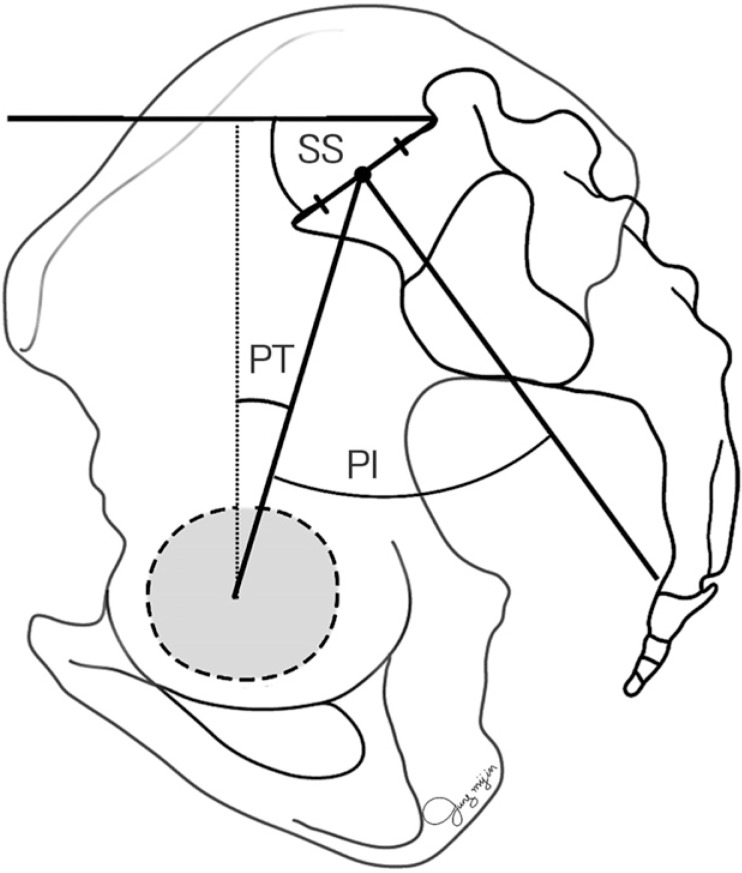
Artistic illustrations depicting the pelvic incidence (PI), sacral slope (SS), and pelvic tilt (PT).
The preoperative DSA, LL, SA, and pelvic parameters415) were measured manually using PACS (Infinitt Healthcare Co., Seoul, Korea). The angular parameters were expressed in degrees.
We used the Brantigan-Steffee classification6) (Table 1) to assess the trabecular bone formation on the 12-month postoperative AP and lateral radiographs. We measured the translational motion and SA of the index segment on the 12-month postoperative flexion-extension radiographs.
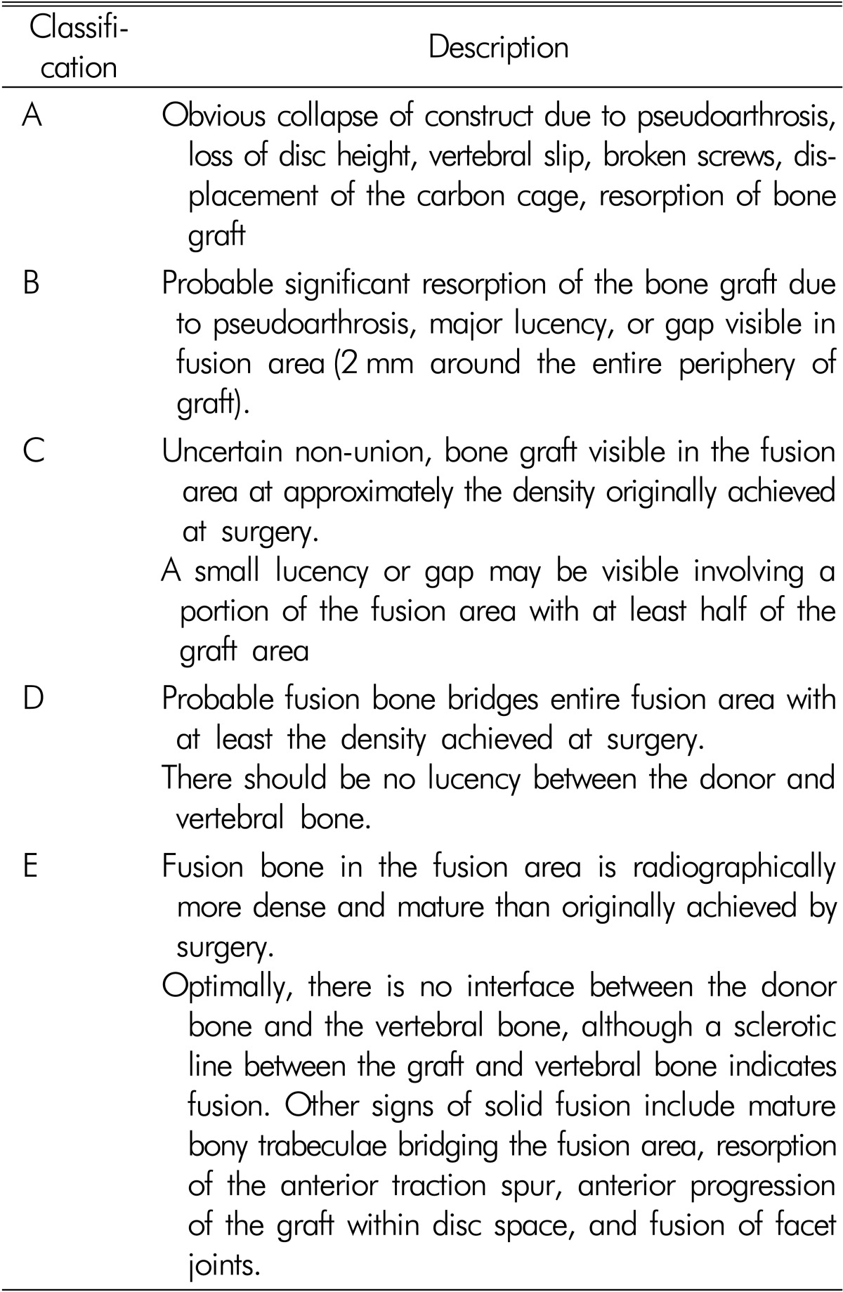
Brantigan-Steffee classification
Fusion was defined as Brantigan-Steffee classification D or E, translation movement less than 2mm and SA less than 5°. Non-union was defined as all conditions except the fusion.
All radiographs were independently interpreted by two spine surgeons (one senior staff and one resident).
3. Surgical Approach
The skin incision was made in the midline of the back, and subperiosteal dissection of the multifidus muscle was performed. After complete bone exposure, total laminectomy or bilateral laminotomy and subtotal or total facetectomies were performed under microscopic visualization. Bone grafts of adequate quantity and quality were obtained from the spinous process, lamina, and enlarged superior and inferior articular processes bilaterally. Then, the ligamentum flavum was resected, and the nerve roots were retracted medially. Complete discectomy was performed, following which the disc space was sequentially distracted, and the endplates were prepared. After completion of central and/or foraminal decompression, two interbody cages filled with morselized local bone chips were placed bilaterally, and morselized local bone chips were packed between the cages. After the placement of interbody cages and autologous bone graft, pedicle screw-rod instrumentation was performed using the conventional technique. Meticulous hemostasis, placement of drain, and layered wound closure were performed serially.
4. Statistical Analysis
Data were expressed as mean±standard deviation in continuous variation or as the number of patients with the percentage in categorical variation. The baseline data, preoperative DSA, LL, SA, and pelvic parameters for fusion and non-union were analyzed using either the chi-square test or Mann-Whitney test. A p<0.05 was considered significant. All statistical analyses were performed using the SPSS version 21.0 (IBM Corporation, Armonk, NY, USA).
RESULTS
A total of 73 patients met the inclusion criteria, of which 59 patients (80.8%) were operated at the L4-L5 level (L4-5 group) and 14 (19.2%) at the L5-S1 level (L5-S1 group). The study included 21 men and 52 women with an average age of 61.8 years (range 42-78 years). The mean duration of followup was 34 months (range 12-102 months). Twenty-two patients had lumbar spinal stenosis without spondylolisthesis, 47 patients had lumbar spinal stenosis with spondylolisthesis, and 4 patients had spondylolisthesis.
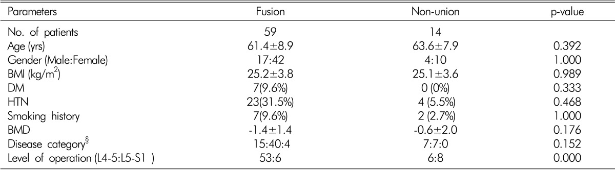
The distribution of age, sex, body mass index (BMI), diabetes mellitus (DM), hypertension (HTN), smoking history, and bone mineral density (BMD) was not significantly different between the two groups (Table 2). According to the diagnosis, L4-5 group had 14 cases of spinal stenosis (23.7%), 41 of spinal stenosis with spondylolisthesis (69.5%), and 4 of spondylolisthesis (6.8%). On the other hand, the L5-S1 group had 8 cases of spinal stenosis (57.1%) and 6 of spinal stenosis with spondylolisthesis (42.9%). The disease category of the two groups was statistically different (p=0.041, Table 2).
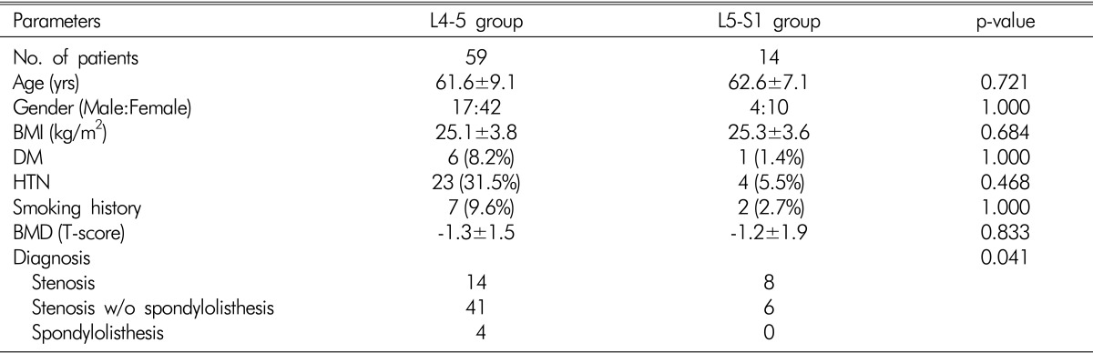
Baseline clinical characteristics of the patients (according to the levels)
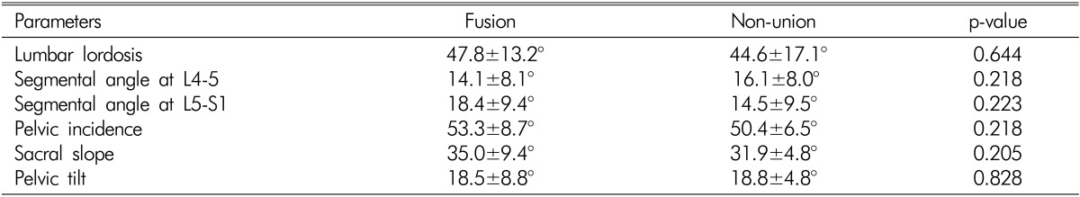
The DSA of the L4-5 group was lower than half that of the L5-S1 group (13.1±8.1° vs. 27.2±6.7°, p<0.001) (Table 3). The SA at the L4-5 level was also lower in the L4-5 group than in the L5-S1 group (13.4±7.9° vs. 18.8±7.6°, p=0.014). However, the SA of L5-S1 was not different between the two groups. LL, PI, and SS were not different between the two groups (Table 3).
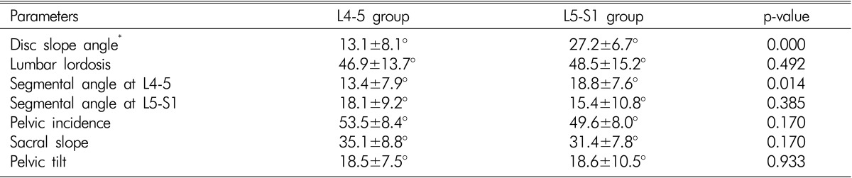
Preoperative radiographic parameters of the L4-5and L5-S1 groups
The radiographic fusion rates at postoperative 12 months were 89.8%(53 of 59) in the L4-5 group and 42.9%(6 of 14) in the L5-S1 group (p<0.001, Table 4.). Fig. 3 and 4 showed the example of the fusion and the non-union.
Comparison of the clinical characteristics of the patients according to radiographic fusion or non-union

The immediate postoperative (A) and 1-year postoperative (B) lateral radiographs of a 59-year-old woman who underwent L4-5 posterior lumbar interbody fusion (PLIF). The 1-year postoperative radiograph (B) showed solid fusion between the vertebral body and the graft.
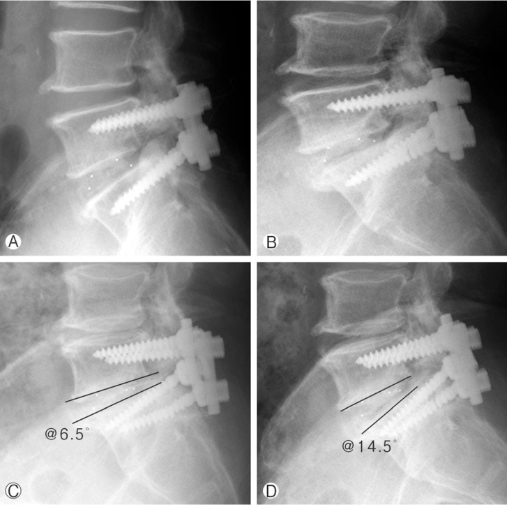
The immediate postoperative lateral radiographs (A) of a 74-year-old man who underwent L5-S1 posterior lumbar interbody fusion (PLIF). The 1-year postoperative lateral radiograph (B) revealed a screw fracture, loss of disc height, and a visible gap in the fusion area. The segmental angle difference was >5° in the fusion segment between the flexion (C) and extension (D) radiographs at 1 year after the surgery.
Among the clinical characteristics of the patients, according to radiographic fusion or non-union, age, sex, BMI, DM, HTN, smoking history, BMD, and disease category were not significant. However, the level of surgery was significant (p<0.001, Table 4). The preoperative LL, SA, and pelvic parameters showed no statistical difference between radiographic fusion and non-union (Table 5). We also evaluated these radiographic parameters in each group according to the fusion status. The preoperative DSA, LL, SA, and pelvic parameters were not statistically different according to the radiographic fusion in both groups (Table 6).
Preoperative radiographic parameters between fusion and non-union
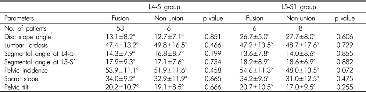
The relationship between presence of fusion and preoperative radiographic parameters in the L4-5 and L5-S1 groups
DISCUSSION
1. Radiographic Fusion Rate
PLIF may be regarded as a more reliable surgical approach for spinal instability than posterolateral fusion because it provides more stable biomechanical conditions in the fusion bed by the axial support and the physiologic lordotic angle with the use of interbody cages9). It also has other advantages such as restoring the disc height, correcting segmental instability, and adequate decompression of the neural elements30). In our institute, most lumbar fusion surgeries for lumbar spinal stenosis or spondylolisthesis have been performed by PLIF rather than posterolateral fusion. However, only a few articles have reported on fusion rates and factors related to single level PLIF. In a study by Aggazzi et al.1), the fusion rate at L4-5 was 96.2% (25 of 26) and at L5-S1, it was 95.2%(20 of 21). Ito et al.11) also reported a fusion rate of 96.4%(80 of 83) at L4-5 and 87.5%(7 of 8) at L5-S1. In contrast, the fusion rate of the L5-S1 group in our study was much lower than that documented in the literature. This low fusion rate in the L5-S1 group may be due to several factors. Our assessment of the fusion rate was based on radiographs, and it was more difficult to determine bony trabeculation at L5-S1 than at L4-5 in both AP and lateral radiographs. The relatively smaller number of patients in the L5-S1 group may also have been inadequate for assessment of the fusion rate. We intended to evaluate the influence of individual levels on the fusion rate. With that in mind, we set up the exclusion criteria, and many cases of L5-S1 were eliminated. The surgical techniques of bone grafting and instrumentation also may have played a role in the lower fusion rate of the L5-S1 group. We used the similar amount of local bone graft at both levels. The amount and quality of the bone graft may have been inadequate for L5-S1. Nevertheless, our results showed an obvious lower fusion rates at the L5-S1 level than at L4-5 with single level PLIF surgery, because we performed the operations with nearly uniform fashion. In addition, no other factors, except the index surgery level, could have affected the fusion rate.
2. Anatomical and Biomechanical Differences
An inferior fusion rate in the L5-S1 group may be due to the anatomical and biomechanical differences between the L4-5 and L5-S1 levels. First, the DSA, which is defined as the angle formed by the disc space and horizontal plane, in the L5-S1 group was two times higher than that in the L4-5 group (27.2±6.7° and 13.1±8.1°, respectively). With an increase in the DSA, the shear force in the disc space also increases. Therefore, this may be a less favorable factor for the fusion rate at the L5-S1 level compared to the L4-5 level. Second, the vertebral compression strength should be considered22). As the lordotic curvature of the lumbar spine begins from the L3 or L4 vertebral body, the compression strength of the lumbosacral articular surface may be lower than that of the L5 vertebral body. As a result, the compression strength exerted on the L5-S1 disc space may be weaker than that exerted on L4-5, and this phenomenon is also disadvantageous for PLIF at L5-S1. The third factor to be considered is the larger range of motion at the L5-S1 segment. In the lumbar spine, flexion and extension motions increase in range from the top to the bottom, and the lumbosacral joint offers more flexion and extension motion than any other lumbar segments321). These facts indicate that translational movement between the L5 vertebra and sacral promontory may occur in larger scale than any other lumbar segments after PLIF. The fourth factor is the facet joint orientation between the L5 and S1 vertebrae, which is in a more coronal plane than other lumbar segments13). This facet joint orientation is very important for the stability of individual segments. However, we performed total or subtotal resection of bilateral facet joints for adequate foraminal decompression. This wide resection of the facet joints was inevitable for relief from compressive symptoms but may be detrimental for segmental stability and successful bone fusion after PLIF. This issue of facet joint resection may be more important for the L5-S1 segment because facet joint strain on L5-S1 is greater than that at other lumbar spine segments2). Finally, the shape of the disc space should be considered. The L5-S1 disc space is conical in shape, with the posterior margin of the disc being narrower than the anterior margin. On the other hand, the disc space in other lumbar spine segments is shaped more like a rectangle. We used fusion cages of the same size and lordotic angle at both levels. Therefore, a contact between the bony end plate and surface of the fusion cage might be less compact and tight at the L5-S1 level than at the L4-5 level. We also experienced some difficulty in packing dense bone graft in the anterior disc space of L5-S1 because of a narrow posterior disc space.
3. Pelvic Parameters and Bone Fusion
We also investigated the effect of preoperative pelvic parameters on bone fusion after PLIF. The pelvic parameters are related to the sagittal balance of human spine and may be altered in certain pathologic conditions such as degenerative or isthmic spondylolisthesis16). Therefore, we hypothesized that differences in pelvic parameters may influence fusion rates after PLIF. Lee et al.14) studied pelvic parameters in the normal Korean population and reported a PI of 52.5±4.5°. According to their result, the values of the two groups in this study were within the normal range for the Korean population. Preoperative PI, SS, and PT were not different between the two groups. Although patients with fusion had higher PI, SS, and PT than the non-union cases in each group, the difference was not significant. Our result shows that preoperative pelvic parameters are not related with a successful bone fusion after single PLIF regardless of the index level of operation. In other words, the sagittal balance of a patient with degenerative lumbar spinal stenosis or spondylolisthesis is not related with radiographic fusion after single level PLIF.
4. Suggestions for Successful Bone Fusion at the L5-S1 Level
As our result showed that the local environment is less favorable for successful bone fusion at the L5-S1 level than other lumbar segments, several technical considerations may be required during the surgery. The first one is the need for more precaution and a delicate procedure for fusion, both of which are achieved thorough denudation of the cartilaginous end plate, compact packing of the bone graft, especially in the anterior disc space, and use of a good quality as well as sufficient amount of bone graft. The second is an optimal instrumentation technique, such as the use of interbody cages that fit the shape of the disc space (lordotic configuration for L5-S1 level), pedicle screws with thicker diameter for S1, and bi-cortical purchase of S1 pedicle screws. The third important consideration is that unless it is absolutely necessary to perform decompression, a bilateral total facetectomy should be avoided.
5. Limitations of Study
There are several limitations to this retrospective study. This study was conducted retrospectively. First, the number of enrolled patients was small, and the distribution of patients in each group was unbalanced. The second limitation was anatomical and related to the ilium in the radiographs. In the lateral radiographs, the L5-S1 disc space was more shrouded by the iliac bone than the L4-5. Therefore, confirming the trabeculation was more difficult in the L5-S1 group than in the L4-5 group, and it was also somewhat difficult to clearly confirm the fusion or non-union state. Third, using the CT scan, the fusion rate could be evaluated more accurately than by radiography7). However, in this study, a CT scan was not available in some cases, and therefore could not be included as an evaluation method. Our data, however, had a uniform disease category and surgical technique using bone grafting and instrumentation.
CONCLUSION
The fusion rate at the L5-S1 level was much inferior to that at the L4-5 after single level PLIF. The anatomical and biomechanical aspects of these two levels are different. The pelvic parameters were not statistically significant between L4-5 and L5-S1 and were unrelated with bone fusion. To attain successful bone fusion at the L5-S1 level with PLIF, one should use good quality and sufficient amount of bone graft, fusion cages that fit well in the disc spaces, and rigid fixation techniques, as well as avoid bilateral total facetectomy, except in inevitable cases.