INTRODUCTION
Dural tearing is not an uncommon complication during lumbar surgery2,12). It mostly can be detected during surgery and repaired easily. However, incidental durotomy without visible dural tear and leakage of cerebrospinal fluid (CSF) can not be readily found intraoperatively. If once incidental durotomy occur, it is able for nerve root to be entrapped. In this article, we report 2 cases of incarceration of spinal nerve root through incidental durotomy and discuss its possible mechanism.
Case Reports
1. Case 1
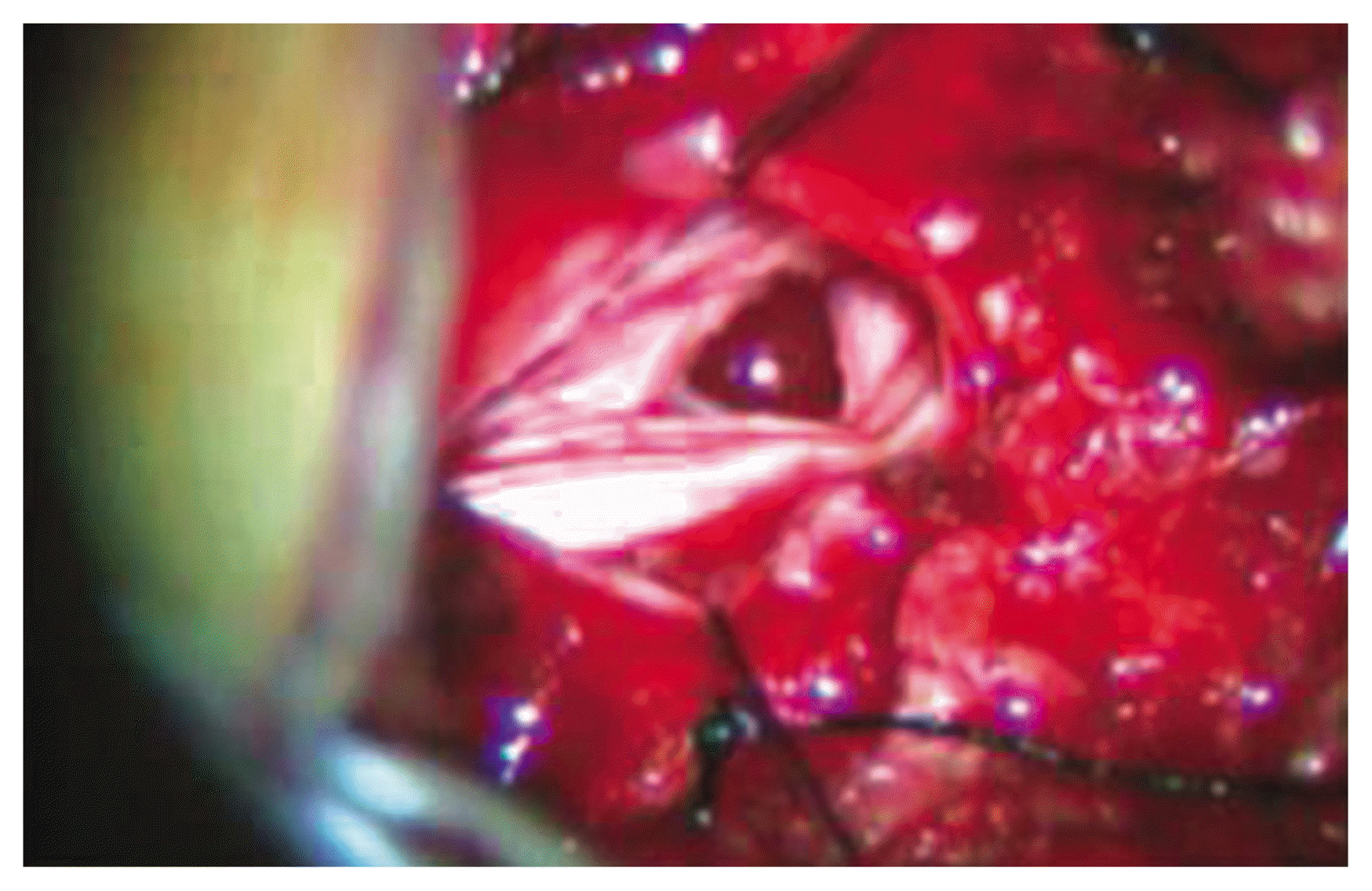
This 43-years-old woman was suffered from progressive left-sided sciatic pain for 6 weeks. Neurological examination demonstrated no motor deficits. Magnetic resonance imaging (MRI) revealed a paracentral disc herniation at the L4–L5 level on the left side. The patient underwent partial hemilaminectomy and discectomy. The herniated disc material was identified and removed. The operative procedure was uneventful. Either dura tearing or CSF leakage did not detected on microscope. The patient improved and no neurological deficits were noted. At over 3 months, sudden onset of severe sciatica and weakness of the left foot was occurred. MRI revealed disc herniation and fibrocystic granulation tissue (Fig. 1). The patient was scheduled for a second operation. The previous laminectomy at L4 was extended. The disc herniation did not recur, but ventral dural tear was seen and nerve root was incarcerated through dura tearing (Figs. 2, 3). After subtotal laminectomy, dorsal dura was opened. The intradural disc was seen in the thecal sac. In addition to removal of intradural disc material, the nerve root was repositioned and dura was repaired. She had prompt recovery soon after the surgery with complete resolution of the pain. And the weakness of the left foot subsided slowly.
2. Case 2
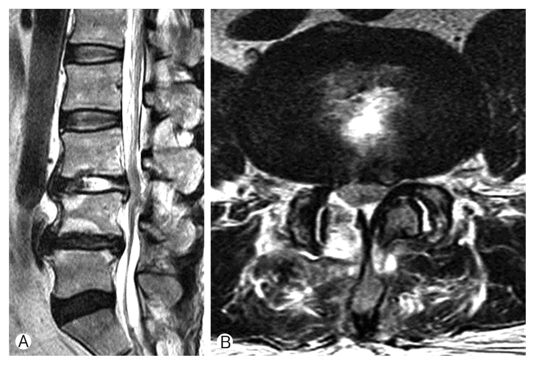
This 61-year-old woman presented with recurrent bouts pain on low back and right leg over the course of 2 months. Neurological examination demonstrated no sensory or motor deficits. MRI revealed a paracentral disc herniation at the L3–L4 level on the right side. She underwent L3–L4 discectomy. After removal of the inferior third of the superior lamina and superior third of the inferior lamina, extruded disc material was extirpated. The operative procedure was uncomplicated on microscope. There was no visible dural tear and no CSF leak. Her symptoms were relieved until the third postoperative day. The patient complained of progressive severe radiating pain. MRI revealed minimal fluid collection at the same disc space without something wrong (Fig. 4). Because of impending cauda equina syndrome, exploration had to be done. At operative field, ventral dural tearing and incarceration of edematous nerve root were noted (Fig. 5). Following midline incision of the dorsal dura, incarcerated nerve root was repositioned and duroplasty was done. Postoperatively, the patient’s sciatica was immediately alleviated and did not recur during 1-year follow-up period.
DISCUSSION
The spinal dura encloses the nerve roots and CSF. When the 2 layers of cranial dura enter the spinal canal, the outer layer blends with the periosteum of the cervical laminae within the canal, and the inner layer joins the arachnoid and becomes the spinal dura5). Inner and outer surfaces of the spinal dura are covered by flattened fibroblasts, and the dense membrane is separated from the periosteum by a narrow epidural space10). Because the spinal dura consists of the only inner membrane of 2 layers, compared with the cranial dura, it may be prone to tear and rupture. According to advances in instrumentation, surgeons get more aggressive attitudes toward spine surgery which could not be performed previously, and are likely to encounter increasing numbers of the dura tear. It is reported that the incidence of iatrogenic dural tears is about 0.3%–13%, but varies according to the type of surgical procedure performed, repeated operation, lesion level, and use of instruments1,3,7,8,13–15). How does extradural incarceration of the spinal nerve root develop, even though no event intraoperatively? The acute pressure of the protruded disc may erode and then thin the dura6,9). The existence of adhesions between the ventral dura and the posterior longitudinal ligament is likely to develop dural thinning and subsequent perforation4,11). This mechanical irritation and adhesion caused by disc inflammatory process may be predisposing factor to make dura thinning. Also, it must be considered intraoperative partial injury of the spinal dura in primary surgery.
To explain injury mechanism of ventral side dura, following theories are worth consideration. Spine surgeons remove ruptured disc particle after retraction of nerve root by using nerve hook or retractor. Ventral side dura layer might be trapped between retractor and dorsal surface of vertebral body, because dura is rolled to medial direction when dura retracted by nerve retractor. That is why caution is needed during nerve root manipulation. Another caution is using of pituitary forceps during removal of disc in disc space. When pituitary forceps go through into disc space, forceps should be closed, because opened forceps could damage to ventral dura during dura retraction.
After intraoperatively partial injury of the spinal dura, neither rupture of the arachnoid membrane and leakage of CSF occur, incidentally increased cerebrospinal pressure (e.g., coughing duration extubation) lead arachnoid membrane to bulge easily due to thin and delicate nature of the arachnoid membrane. The rupture of the arachnoid membrane causes leakage of the CSF postoperatively, and extradural herniation of nerve root occurs through this opening gradually. At this time, herniated nerve root has action of check valve and begins to swell and be incarcerated. If once swelling and incarceration of nerve root, patient suddenly complains of severe sciatica and motor weakness. In our case, direction of dura opening is all ventral to the dural sac. The authors think that incidental durotomy has to do with operative procedure. Pituitary forceps, root retractor and sharp suction tip worn off by drill are able to cause intraoperatively partial injury of the dura.
CONCLUSION
Incidental durotomy should be respected for its potential associated complications and medicolegal implications. The surgeons must be care of avoiding dura tear. When patient undertakes uncommon radiating pain and neurological deficit after lumbar discectomy, nerve root incarceration must be taken into consideration in the differential diagnosis.