INTRODUCTION
Spinal fusion is a popular management option in the management of degenerative conditions of the lumbar spine4). Since it was first described by Briggs and Milligan in 1944, the posterior lumbar interbody fusion (PLIF) has some distinct theoretical advantages over posterolateral techniques as a fusion strategy4). The benefits of the PLIF are securing the fixation of vertebral body, maintaining the normal intervertebral space and supporting the anterior column in charge of 80% of weight-bearing out of the vertebral column, thus providing satisfactory bone fusion while maintaining biomechanical stability22,26-28). Various kinds of spinal implant cage devices have been designed to provide a relatively simple and effective technique for implementing PLIF and to improve fusion rates by acting as a structural support while biological fusion occurs8,14). Recently several authors have reported that the cage geometry, such as rectangular, trapezoid or cylindrical, has a significant impact on the alignment of the lumbar spine after instrumented PLIF9,13,20). These authors considered wedgeshaped, trapezoid cages significantly increase segmental lordosis, enhancing lumbar lordosis, and therefore should be preferred for restoring sagittal alignment in instrumented PLIF procedures9,13).
We have utilized rectangular cages for PLIF since 1995. In the present study we investigated whether the lumbar sagittal alignment can be obtained within normal range in patients who have undergone PLIF with stand-alone cages, which have no intrinsic contour to induce lordosis and assessed not only the firm bone fusion but also the clinical results of those patients. Here, we had an opportunity to review the mid-term follow-up outcomes from patients who underwent placement of rectangular stand-alone cages by a single, independent surgeon.
MATERIALS AND METHODS
Patient Selection
Patients with back pain with or without radiating pain who underwent PLIF with stand-alone rectangular cages between 1996 and 2004 have reviewed retrospectively. Radiographic and clinical follow-up of patients was reviewed in 33 patients who were followed-up at least 4 years after surgery. Thirteen patients had failed back surgery syndrome after primary disc surgery, 9 had herniated intervertebral disc, 7 had degenerative disc disease (DDD), 3 with spinal stenosis, and 1 had spondylolisthesis. The cages used including Ogival Interbody Cage (OIC), Carbon Cage (CC), CH Cage, Poly-Ether-Ether-Ketone Cage (PEEK).
A total 33 patients in age range of 22 to 74 years with the mean of 46.2 years enrolled into the study. The summary of patient demographic data is listed in Table 1. Surgery was performed on patients who mainly had symptomatic degenerative disc disease showed that definite low signal intensity on T2 MRI, definite decrease in the intervertebral disc height in one or two contiguous lumbar levels, unresponsive to conservative treatment and no greater than Grade I spondylolisthesis. Patients were excluded from the study if they had any symptomatic disc disease at a level other than L4-L5 or L5-S1 or a severe medical condition.
Surgical Procedure
All surgical procedures were carried out by a single spine surgeon. Patients underwent total laminectomy and medial facetectomy to visualize thecal sac and nerve roots to make sure decompression is sufficient. And bilateral discectomy was held using shaver, then posterior lumbar interbody arthrodesis with two cage devices were implanted at either the L4-L5 or L5-S1 lumbar interspace. Four kinds of rectangular cages were used in our series namely Ogival Interbody Cage (OIC: Stryker Howmedica Osteonics, Mahwah NJ, USA), Carbon fiber (CC: De Puy-Acro med Co., Raynham, MA, USA), CH Cage (Spine-Tech, Minneapolis, MN, USA), Poly-Ether-Ether-Ketone (PEEK, Stryker Howmedica Osteonics, Mahwah NJ, USA) cage. These cages were without any lordotic angle such as 0 degree and have little or no intrinsic ability to induce a lordotic contour. The chamber of cages was filled with autologous cancellous bone obtained from the lamina or iliac crest except 2 cases used allograft bone chip. Every patient had orthothic device for minimum 2 months. The results from all the studies were pooled and analyzed independently to define the effects of the surgical technique on the surgical outcome, hospital stay, and the mid-term clinical and radiographic outcomes.
Assessment of Clinical and Radiographic Outcome
Through pre- and post-operative direct evaluation at 1, 6, 12 and 24 months on their hospital visit or a telephone survey, the severity of low back or leg pain was evaluated by a Visual Analogue Scale (VAS) and the clinical outcomes were examined by an Odom's criteria.
Plain radiographs were measured and reviewed by single spine surgeon at pre- and immediate post-operative state, postoperative one year, two years and after in alternate years. Intervertebral height was measured at the mid-point of both lines which are connected from anterior to posterior end plates of upper and lower vertebral bodies on the lateral plain radiographs, total lumbar lordosis was measured from the bottom of T12 to the bottom of L5 as described by Cobb35). The degree of segmental lordosis at the site of surgery was measured from the lower endplate of the upper segment to the upper endplate of the lower segment. Using the simple lumbar lateral X-ray with flexion-extension view, stability and the status of fusion was assessed. Thin-cut computed tomography scans with sagittal and coronal reconstructions through the fusion construct were obtained as necessary. Fusion was defined when all the conditions below were fulfilled. Bridging bone connecting the adjacent vertebral bodies either through the implants or around the implants, <5° of angular motion, ≤3mm of translation, and an absence of radiolucent lines around >50% of either implant. Secondary lumbar surgical procedures performed subsequent to the index operation because of a suspected nonunion, regardless of the radiographic findings, were classified as second surgery failures and fusion failures5).
Statistical Analysis
All statistical analysis was carried out using SPSS (version 17.0, SPSS Inc., Chicago, IL). The changes in preoperative and postoperative radiological findings were analyzed using the paired t-test. The Mann-Whitney test was used. Statistical significance was determined when p values were less than 0.05.
RESULTS
Patient Demographics
Thirty-three patients who were followed up at least four years, twelve patients (36.3%) were followed more than 8 years. Average surgery time was 236.8±53.1 minutes, average intraoperative blood loss was 334.8±292.2 mL, and average hospital length of stay was 10.8±3.3days. All of the patients used autologous bone chip. Further, thirty (90.9%) patients of them used autologous iliac bone and 3 (9.1%) used autologous and allograft mixed bone chip.
Three patients (9.1%) experienced perioperative complications including dura tearing which occurred two times, however, there were no CSF leakages or no meningitis post-surgery. One patient had aggravation of stenosis at L3-4 level that needed operation. There were no other clinical complications including neurologic deficit, infection, hematoma formation, hardware failure or cases of re-operations were observed during the follow-up period.
Clinical Outcomes
The mean score on the VAS of back pain found to be improved from 8.0±2.9 points during the preoperative period to 3.4±3.0 points at one year postoperative and decreased to 2.1±3.0 and 3.5±2.7 at postoperative 4 and more than 8 years, respectively. The VAS of sciatica was reduced from 7.1±3.1 at preoperative to 2.5±2.8 post 1 year, and it was reduced to 1.5±2.8 and 3.1±2.6 at the same periods, respectively. At their last follow-up, about 94% of patients showed excellent or good outcomes on Odom's criteria (Table 2).
Radiographic Outcomes
The results on the change of the intervertebral disc height, segmental lordosis and total lordosis are also listed in Table 3. The mean intervertebral disc height was 8.2±1.4mm before surgery and it was increased to 10.7±1.2mm at postoperative and was decreased to 8.7±1.9mm by 2 years and it was stabilized to 8.3±1.8, 8.1±2.1 and 8.7±1.9mm at 4, 6 and more than 8 years follow-up visit. It was modestly decreased on the final visit compared with postoperative state. The angle of segmental lordosis at the neutral position was increased from 12.1±4.0° before surgery to 14.1±4.4° by postoperative 1 year. The angle was maintained for two years with no significant difference (p=0.317), but it reduced at 4 year follow up (p=0.072). The angulation of total lumbar lordosis also changed from 32.8±10.2° to 35.3±8.9° at the same period (p=0.200). But there was not statistically significant difference.
Plain X-rays were analyzed for confirming solid fusion at the out-patient clinic during follow-up period. A total of 30 patients with obvious trabecular bridging on the plain x-ray and the fusion rate was 87.9% at 1 year follow-up. The fusion rates were 91% at 4 years follow-up.
There was no significant difference of clinical results between 4 types of cages. There was significant difference between 4 types of cages in radiological outcome. CC had excellent radiological outcome in disk height, segmental lordosis and total lordosis during 4 years of follow-up. Other cages also seemed to improve in radiologic outcome but we could not find statistical significant difference (Table 4).
Illustrative Case
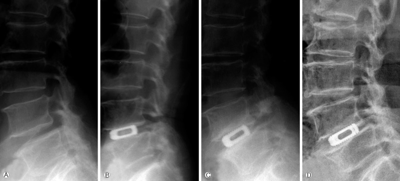
Case I
Fourty nine years old male who complained with left leg pain had disc herniation at lumbar 4-5 level and underwent PLIF with CC cage (Fig. 1). Height of disc space was 9.5mm before surgery and height rise up to 12 mm. After a year, height slightly decreased to 11 mm and the height maintained in 10 mm during 7 years of follow-up. Segmental lordosis before surgery was 13.5° which merely changed after surgery. But after 7 years it decreased to 7°. Total lordosis before surgery was measured as 42.9° which decreased in 33° after surgery and changed minimally till 7 years of follow-up. His VAS score decreased from 9 to 2 and excellent in Odom's criteria.
Case II
Sixty one year-old lady suffered from her both buttock and leg pain for 20 years. Her symptom aggravated since 2 years before admission, and diagnosed to have spinal stenosis at lumbar 4-5 level. She underwent discectomy with OIC cage insertion (Fig. 2). The Disc height was improved from 8.3mm to 11 mm after surgery. After a year, height slightly decreased to 9 mm with suspicion of subsidence and the height maintained in 8 mm during 8 years of follow-up but had Grade I spondylolisthesis. Segmental lordosis before surgery was 15° which have decreased to 13.3° after surgery and increased to 18° in 8 years of follow-up. Total lordosis before surgery was measured as 44.8° which decreased in 42° after surgery and gradually increased to 50° after 8 years. Her VAS score before surgery was 8 for back pain and 5 for leg pain which decreased to score 0 for both. With confidence, excellent result in Odom's criteria.
Discussion
Lumbar interbody fusion provides several theoretical advantages over other fusion techniques28,31) including biomechanical stability with a higher fusion rate33) and can create the restoration of disc height and the sagittal balance26). Furthermore, since stand-alone PLIF needs less muscle retraction leading to less complications and post-operative pain10). As Lumbar interbody fusion inserted posteriorly, the spinal canal can be easily explored. Furthermore, the use of locally derived bone obviates the need to harvest iliac bone or the amount of bone required for the graft27). The disadvantages include interbody fusion Problems such as collapse, slippage, and graft migration in 3 to 10% of cases in major studies1,16). Particularly, in PLIF, dural and nerve root manipulations represents as a risk of this procedure11,15,17). However, taken the above merits into account PLIF is still considered as a primary choice.
Clinical Outcome
The clinical outcome after PLIF can vary widely based on the selection criteria. As we chose to use VAS and Odom's criteria, the significant reduction of VAS was achieved at 1 year after surgery and the reduction of VAS lasted to follow-up periods even if it was found to be increased. These results are in agreement with a 2 year follow-up study19). The analysis of Odom's criteria represents clinical outcomes indicating the maintenance of the successful clinical results with mid-term follow-up.
Fusion Outcome
Several studies indicate, the bone fusion rate of other interbody fusion methods are more than 90%3,16,22,24,25,32). In the present study, the fusion rate was 91% at 4 years follow-up and found to be constant during subsequent years of follow-up. The fusion rate of stand-alone cage insertion may be lower than followed screw fixation. Compared with other stand-alone PLIF series (85.2% to 86%)29,36), the result was better in this series but lower than pedicle screw fixation group (91.1%)12).
The fusion was confirmed by plain x-ray or CT scan. The flexion-extension film showed stability in all patients. As described by Fraser26), it is difficult to specifically ascertain the fusion rate in radiologic findings and a simple comparison for fusion rate is impossible without any criteria. Therefore, in this study, we used the criteria suggested by Burkus6).
Disc Height, Subsidence and Lordosis
Subsidence or spondylolisthesis with instability is the most common matter of concern after PLIF with stand-alone cage. In the present study there were differential subsidence in most cases but the subsidence was progressive up to 2 years post-surgery. The intervertebral disc height was reduced by approximately 6% after 4 years of surgery. However, the rate of subsidence was decreased to 2% per year. The data obtained in our studies showed a relatively early high subsidence rate and a late low subsidence rate in long-term follow up. The cage seemed to maintain the intervertebral disc height as well in the mid-term follow-up.
Interestingly, a lower subsidence rate (about 6.5%) for 2 year follow-up was also reported19). This difference may be due to a difference between a rectangular cage and an expandable cage.
The physiological curve of the spine is related to the distribution of optimal weights loaded onto the spine and the loss of physiological curve in the lumbar spine is attributed to back pain1,11,15,17,18,37). As suggested by Wambolt and Spencer2,7,18,21,34), the destruction of lordosis results from the decrease in intervertebral disc height and interspinous ligament damage due to degenerative changes.
The segmental angle of lordosis was improved from 12.1±4.0° to 10.3±5.8° by postoperative 4 year. The angle was maintained for four years with no significant difference (p=0.179). In spite of using no lordotic angled cages in all cases, segmental lordosis was developed close to physiological lordosis, indicating subsidence of posterior vertebral body has played an important role in making the angle. However, at the forth year follow-up segmental angulation was reduced significantly.
Total lumbar lordosis found to be changed from 32.8±10.2° to 36.0±8.1° at the four years of follow-up. It may be suggested that the segmental angle recovery of the lower lumbar spine is not the only factor in deciding the total lumbar lordosis recovery, however, it is important in the development of the facet joint degeneration, ligament hypertrophy, and back muscle atrophy. The significance of these factors was not evident in this study19).
Complications
Complications associated with PLIF can be serious, especially the neurological deficits often related to excessive retraction of the nerve roots or the dural sac. According to the various reports, these complications occur in 4 to 10% of patients16,22,31). As reported by Kuslich et al.,22) the complication of cage migration was observed in 3% of the patients. Additionally, increasing numbers of cage migration is reported when stand-alone cages were used. Though repeated migration of cage after posterior lumbar interbody fusion has been reported23), the rate of cage migration in patients with no posterior instrumentation was significantly higher compared with the rate in those with posterior instrumentation (16.7% vs. 0%)30). We have observed relatively low of perioperative complications including hardware problems after PLIF using stand-alone cages, which could be due to low rate of cage retropulsion, may be related to the threaded cage appearance.
Study Limitation
There are several limitations in this study which include is only a retrospective review in which preoperative functional data on the patients are not available and lack of adequate clinical follow-up. The correlation between plain radiographic fusion and actual fusion was also not well established as described earlier10,30).
CONCLUSION
We have investigated for the first time, the safety and efficacy of the rectangular cage in the degenerative lumbar spinal disorders. The use of rectangular stand-alone cages for PLIF resulted in a various degree of subsidence. However, the progress of subsidence was halted as fusion progresses despite of using no lordotic angled cages. Segmental lumbar lordosis was naturally developed close to physiological lordosis suggesting that subsidence of posterior vertebral body may have played an important role in making the angle. Results of this study demonstrate very low complications of the cage during the follow-up periods, high functional stability, improved clinical outcomes in patients with degenerative lumbar disc disease.