Type I Chiari Malformation Without Concomitant Bony Instability: Assessment of Different Surgical Procedures and Outcomes in 73 Patients
Article information

Abstract
Objective
Posterior fossa decompression is the treatment of choice in type 1 Chiari malformation (CM-1) without bony instability. Although surgical fixation has been recommended by a few authors recently, comparative studies to evaluate these treatment strategies using objective outcome tools are lacking.
Methods
Seventy-three patients with pure CM-1 (posterior fossa bony decompression [PFBD], n = 21; posterior fossa bony and dural decompression [PFBDD], n = 40; and posterior fixation [PF], n = 12) underwent a postoperative outcome assessment using Chicago Chiari Outcome Score (CCOS). Logistic regression analysis detected predictors of an unfavorable outcome.
Results
Minimally symptomatic patients generally underwent a PFBD while most of the clinically severe patients underwent a PFBDD (p = 0.049). The mean CCOS score at discharge was highest in the PF (12.0 ± 1.41) and lowest in PFBDD group (10.98 ± 1.73, p = 0.087). Patients with minimal preoperative clinical disease severity (adjusted odds ratio [AOR], 4.58; 95% confidence interval [CI], 1.29–16.31) and PFBDD (AOR, 7.56; 95% CI, 1.70–33.68) represented risks for an unfavorable short-term postoperative outcome. Though long-term outcomes (CCOS) did not differ among the 3 groups (p = 0.615), PFBD group showed the best long-term improvements (mean follow-up CCOS, 13.71 ± 0.95), PFBDD group improved to a comparable degree despite a poorer short-term outcome while PF had the lowest scores. Late deteriorations (n = 3, 4.1%) occurred in the PFBDD group.
Conclusion
Minimally symptomatic patients and PFBDD predict a poor short-term postoperative outcome. PFBD appears to be a durable procedure while PFBDD group is marred by complications and late deteriorations. PF does not provide any better results than posterior fossa decompression alone in the long run.
INTRODUCTION
Type 1 Chiari malformation (CM-1) is characterized by a caudal displacement of the cerebellar tonsils due to an overcrowded posterior fossa [1,2]. This often leads to a true or a functional blockade of cerebrospinal fluid (CSF) circulation across the foramen magnum (FM) leading to a plethora of symptomatology. Nearly two-thirds of such patients develop syringomyelia [2-4]. CM-1 may or may not be associated with an associated bony instability of this region like atlantoaxial dislocation (AAD) or basilar invagination (BI) [5-7]. The treatment principle of CM-1 is to re-establish the altered CSF flow across the FM and addressal of the bony instability is usually sufficient for this purpose in cases accompanied by an AAD or BI [5-7]. On the other hand, pure CM-1, that exists without any associated bony instability, is generally treated by posterior fossa decompression (PFD) [1-4]. There is some controversy in the literature regarding the extent of PFD. Both bony decompression alone (posterior fossa bony decompression, PFBD) and a combined bony and dural decompression (posterior fossa bony and dural decompression, PFBDD) are widely practiced [6-8]. In a meta-analysis that compared the 2 techniques, both procedures were associated with similar rates of lack of significant postoperative clinical improvement [9]. However, PFBDD was significantly less likely to lead to reoperations from nonimprovement, albeit with a higher rate of CSF related complications. Lin et al. [10], however, noted that PFBDD led to significantly higher rates of clinical improvement in the presence of syringomyelia (p = 0.007), not in its absence, and definitely at the cost of a higher incidence of CSF leak and related complications.
More recently, Goel et al. [11-13] have proposed a policy of uniform posterior fixation (PF) in CM-1 even without an associated obvious bony instability. He has hypothesized that all CM-1 represent a manifestation of craniovertebral junction (CVJ) instability and PFD may lead to a paradoxical clinical worsening. Nevertheless, surgical fixation is not the first-choice treatment as yet [1,14-16], and in particular, a recent study by Salunke et al. [17,18] examined this strategy and failed to document any advantage of this approach over the conventional PFD. However, neither study made a comparative assessment of the 3 surgical procedures in the same data set. Recently, objective outcome assessment tools have been promoted to evaluate outcomes following CM-1 decompression [19]. Therefore, our study is an attempt to compare the short- and long-term effectiveness of different surgical procedures for pure CMs using the CCOS system [20,21].
MATERIALS AND METHODS
1. Patient Population and Clinical Assessment
We retrospectively studied 89 consecutive patients with CM-1 operated at our institute between January 2013 to June 2019. The inclusion criteria were: (1) patients without radiological evidence of AAD or BI on preoperative radiology; (2) operated for the first time at our institute. The criteria used for defining AAD and BI and exclusion of these cases were as follows: AAD was defined as an increased atlantodental interval (> 4 mm in children and > 2.5 mm in adults); and BI was defined as location of the tip of the odontoid above the McRae line, the Wackeheim clival canal line and > 3 mm above the Chamberlain line or > 4.5 mm above the McGregor line on the sagittal plane (i.e., extension of the odontoid tip satisfied all the known radiological criteria). The latter was often associated with AAD and described as group A BI by Goel [22]. Our study included a few patients of CM-1 with basilar impression or the so-called type B BI. This bony anomaly is a common companion of CM-1 and contributes to the small posterior fossa in these patients. Here, the odontoid process maintained its relationship with the anterior arch of the atlas and the clivus, thus remaining below the McRae and Wackenheim lines but the tip of the odontoid extended beyond the limit on Chamberlain and McGregor lines. Figs. 1 and 2 are 2 representative cases from our series showing the bony anatomy associated with CM-1. Out of the 89 patients satisfying our inclusion criteria, 16 patients were excluded due to inadequate follow-up information. Therefore, 73 patients (mean age, 26 years; range, 9-60 years; male:female = 48:25) were finally analyzed.
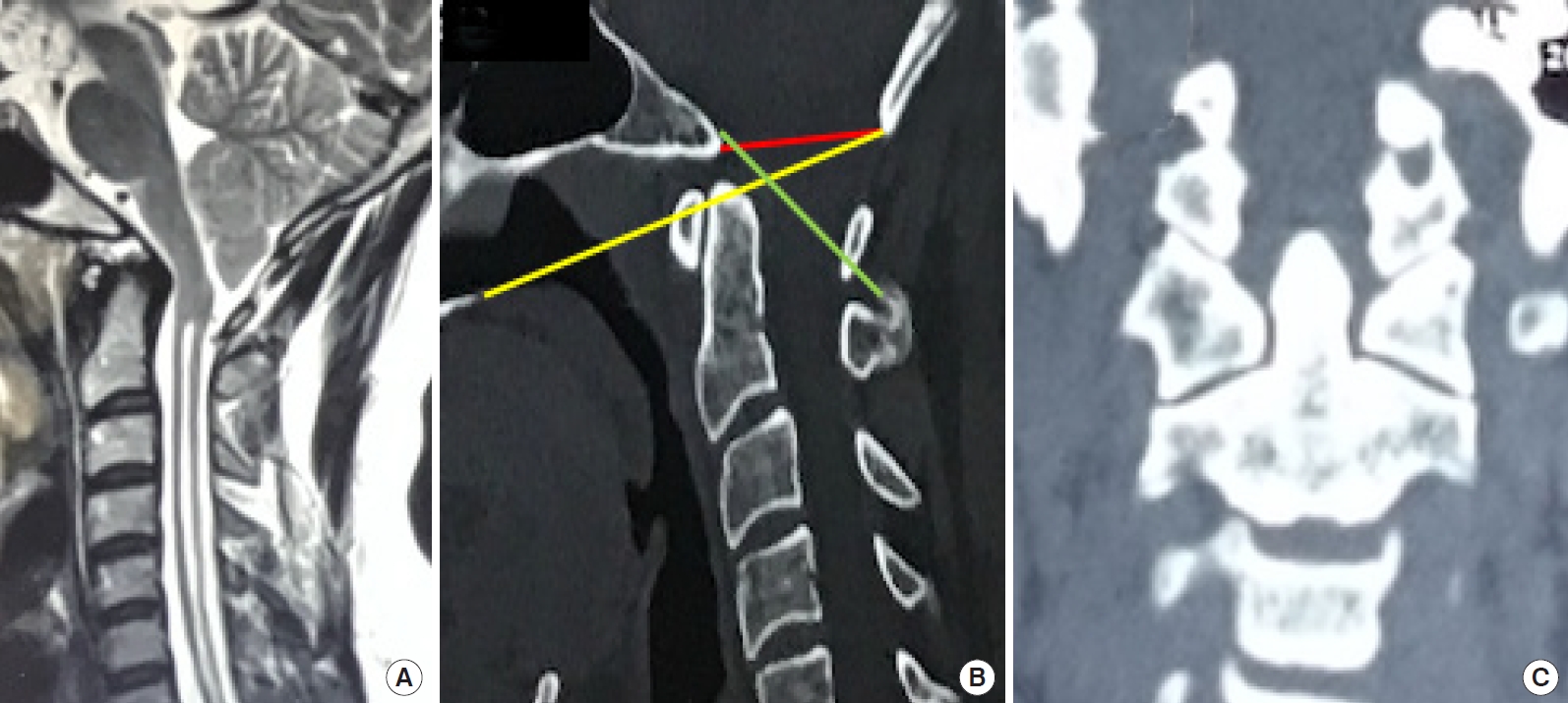
(A) A set of images of a patient with Chiari malformation type 1 with a normal bony anatomy. Sagittal section of the magnetic resonance imaging of cervical spine shows a cervical syrinx, tonsillar displacement below the foramen magnum reaching just above the posterior arch of atlas. On computed tomography evaluation, there are no abnormal bony fusions (B, C) and the odontoid tip is not extending more than 3 mm from the Chamberlain line (yellow) and lying below the McRae (red) and Wackenheim line (green).
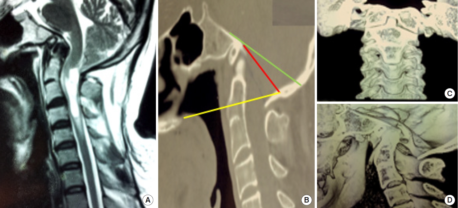
(A) A set of images of another patient with Chiari malformation type 1 with an abnormal bony anatomy. Sagittal section of the magnetic resonance imaging of cervical spine shows a cervical syrinx, tonsillar displacement below the foramen magnum, and ventral encroachment of the medulla by the retroverted odontoid. (B–D) On computed tomography evaluation, there was assimilation of atlas, C2–3 fusion, platybasia with a retroverted odontoid. The odontoid tip is extending more than 3 mm from the Chamberlain line (yellow) but lying below the McRae (red) and Wackenheim line (green), suggesting a basilar impression or type B basilar invagination. In the panel B, the opisthion has been considered to be the point where the 2 cortices of the occipital squama join in view of assimilation of the atlas.
Clinically, we divided the patients into 2 categories, namely ‘minimally symptomatic CM-1’ and ‘clinically severe CM-1.’ The former category comprised patients with headache or neck pain with or without mild paresthesia that was not bothersome (n = 26, 35.6%). On the other hand, the presence of severe paresthesia, motor symptoms (myelopathy), atrophy of the hands with or without dissociative anesthesia were considered as clinically severe disease (n = 47, 64.4%).
2. Neuroimaging Evaluation
All patients underwent magnetic resonance imaging (MRI) of the cervical spine including the cervicomedullary junction. MRI images included one scout image of the entire spine and cranium, apart from the detailed sections of the cervical spine and the posterior fossa to detect an associated syrinx or a spinal curvature anomaly, if any. Radiological findings noted were; extent of tonsillar descent (below the McRae line, expressed in millimeters as well as with respect to the posterior element of the atlas or the axis), presence of syringomyelia (vertical extent), the pB-C2 distance (perpendicular distance between the tip of the odontoid from the line joining the tip of the clivus to the postero-inferior point of the C2 vertebral body), distance between the posterior part of the odontoid and opisthion, craniocervical angle and hydrocephalus. Similar to the clinical categorization, we divided the radiologically perceived severity of the disease into: radiologically mild disease (no syrinx or limited syrinx [cervical/cervicodorsal syrinx not extending to lower thoracic level i.e., below T4 vertebral level] with a tonsillar descent not beyond lower border of C1) and radiologically severe disease (either an extensive syrinx or tonsillar descent upto C2 or below or satisfying both conditions). Extensive syrinx was categorized as: holocord syrinx, distal (below T4) dorsal syrinx, lumbar syrinx, or cervicodorsal syrinx extending below the T4 vertebra. We modified our previously published classification (4 types) of tonsillar descent into 2 types [16].
Additionally, all patients underwent dynamic plain skiagrams or computed tomogram of the cervical spine to assess the bony anatomy.
3. Surgical Treatment
Our patients underwent 2 sets of procedures: the PFD only (n = 61, 83.6%) or PF (n = 12, 16.4%). Bony PFD (n = 21, 28.8%) was accompanied by a division of the dural band in all cases. Augmentation duraplasty was generally added to the bony decompression (PFBDD, n = 40, 54.8%) when there was a syrinx associated with CM-1 (a strategy validated by Lin et al) [10]. However, a few patients underwent PFD even with a localized syrinx and some underwent PFBDD even in the absence of syrinx. For duraplasty, the materials used were artificial dura (n = 6), and locally available fascia in the rest (n = 34). Very few underwent additional intradural procedures like arachnoid lysis and tonsillar shrinkage (n = 12). Surgical fixation as a primary treatment was purely based on the surgeon’s choice and based on the recent publications [12,23-25]. In 9 of these patients, a bony decompression (removal of the posterior rim of FM) was also added.
4. Outcome Assessment
At discharge as well as at the last available follow-up, both clinical and radiological assessments were carried out. Clinical evaluation included gestalt questionnaire and CCOS [19]. The outcome was dichotomized as per a cutoff suggested by Hekman et al. into favorable outcome (scores 11–16) and unfavorable outcome (scores 4–10) [26,27]. This score was used at the time of discharge as well as at last follow-up visit/interview.
As per this scoring system, all but 1 patient had follow-up CCOS score of 11 or more. Therefore, we used a new matrix to estimate the long-term outcomes using the difference between follow-up scores and the discharge scores for every patient. The median value of this score difference (value of 2 in this study) was used to dichotomize the patients into: marked improvement (difference of ≥ 2) or minimal/ nonimprovement (difference of less than 2).
Only those who did not report any satisfactory improvement or had a clinical worsening following surgery underwent a follow-up MRI. The parameters noted were: reduction in the extent of the syrinx, patency of the neo-cisternal magna, changes in the pB-C2 distance (on MRI), or iatrogenic C1/2 joint dislocation (antero-posterior or supero-inferior or rotational) on postoperative computed tomography scan of the CVJ [19,28].
5. Statistical Analysis
Normality of data was examined and normally distributed data were presented as mean ± standard deviation whereas nonnormal data were presented as median (interquartile range). For the continuous data, a comparison between the 2 groups was done using the independent t-test or the Mann-Whitney U-test. For a comparison of mean among the 3 groups, One-way analysis of variance or Kruskal-Wallis test was used. Categorical data were represented as frequencies and compared using χ2 test/Fisher exact test. For the assessment of predictors of the outcome, a binary logistic regression analysis was applied and uni as well as multivariate analyses were performed. IBM SPSS Statistics ver. 22.0 (IBM Co., Armonk, NY, USA) was used for data analysis.
RESULTS
1. Clinicoradiological Findings
Table 1 summarizes the clinical and radiological findings of our study. Males (p = 0.007) and younger adults (18–40 years) were more significantly affected (p ≤ 0.001). Majority of the patients presented with symptoms of more than 1-year duration (n = 55, 75.3%; mean, 30.2 months; range, 1–146 months). The most common symptom was sensory paresthesia with a variable degree of sensory loss (n = 52, 71.2%) followed by pain at the nape of neck (n = 50, 68.5 %), out of which 12 patients also complained of headache. Interestingly, only 8 (11%) patients had urinary symptoms. Fifty-one patients (69.9%) had motor weakness either as hand grip weakness or as limb weakness. Forty-seven patients (64.4%) had some signs of myelopathy like hypertonia, weakness, hyperreflexia, etc. Frank atrophy of the thenar/hypothenar muscles was seen in 9 patients (12.3%), although a far more number of patients complained of weakness. Ten patients (13.7%) had lower cranial nerve signs while a similar percentage of patients had subtle cerebellar signs. There was a significant difference in the clinical disease severity amongst the patients in the different treatment arms (p = 0.049). The group who underwent a PFBDD had a significantly more severe disease while patients undergoing PFBD were mainly minimally symptomatic (Table 2).
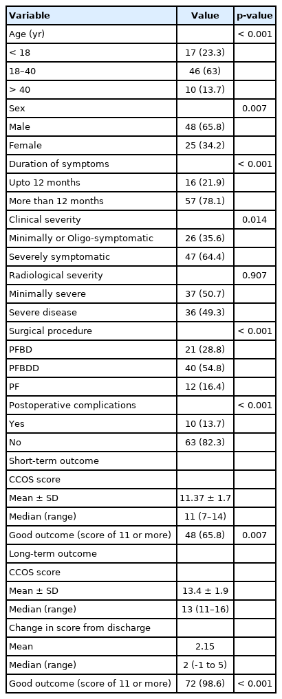
Baseline clinicoradiological findings and surgical outcome results of our study
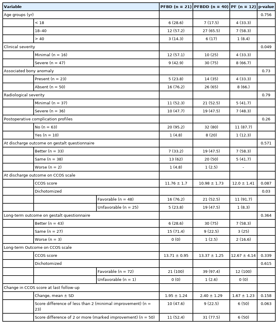
Comparison of clinicoradiological findings and surgical outcomes of the 3 procedures employed in our study
Average displacement of the tonsil was 10.44 ± 5.3 mm (range, 6–30 mm). While 44 patients (60.3%) had tonsillar decent upto the C1 arch, 29 had tonsillar decent below the C1 arch (39.7%). Out of the 73 patients, 65 (89%) had a syrinx. Thirty-three patients (45.2%) had cervicodorsal syrinx, constituting the most common type of syrinx in our study. Using our criteria, 37 patients had extensive syrinx in our series (50.7%) and 28 patients had a limited syrinx (38.4%).
Twenty-three of our patients (31.5%) had an associated bony anomaly out of which occipitalization of the atlas was seen in 18 patients (24.7%) while C2–3 fusion was present in 12 patients (16.4%). Nine patients (13.2%) had basilar impression (type BBI) with platybasia. The distribution of these bony anomalies did not differ significantly among the 3 groups (p = 0.73). The incidence of these anomalies in the 3 groups was as follows: 23.8% (n = 5 of 21) in PFD group, 35% (n = 14 of 40) in the PFBDD group, and 33.3% (n = 4 of 12) in the PF group. The individual incidences of the 3 bony anomalies i.e., assimilation of atlas, C2–3 fusion, and basilar impression with platybasia also did not vary significantly among the 3 groups (Table 2). The mean pB-C2 distance in our study was 8.4 ± 2.8 mm (range, 4.5–16.3). The radiological severity as well as bony anomalies were insignificantly distributed amongst the 3 groups (Table 2).
2. Short-term Postsurgical Outcome
There was no mortality following the primary surgery but there were 10 patients (13.7%) with postoperative complications. Motor worsening was the commonest complication after surgery (n = 4, 5.5%). Two of these patients had a pre-existing minor motor weakness. With respect to the bony anatomy, 2 of them had assimilation of atlas with C2/3 fusion and basilar impression with platybasia. None of these patients had a demonstrable pre- or postoperative instability. Two of them had undergone a PFBDD, one patient each had undergone PFBD and PF respectively. Three patients gradually attained the preoperative power on their own while a salvage PF was performed in one patient (patient No. 1, Table 3). Four patients (5.5%) developed transient CSF-leak related complications out of which 1 patient developed a surgical site infection. Postoperative worsened lower cranial nerve dysfunction was noted in 2 patients (2.7%). While the deficit was transient in one, another patient required a tracheostomy for airway protection. This patient had undergone a PFBDD. She gradually recovered and could be discharged with a CCOS score of 10 which improved to a score of 12 at last follow-up (5 years). Although the number of complications did not differ significantly among the 3 groups (p = 0.26), 80% (n = 8) of the complications occurred in the PFBDD group as depicted in Table 2.

Details of the patients requiring a resurgery with surgical fixation for clinical deterioration in this series
On the Gestalt questionnaire, 33 patients (45.2%) reported an improvement, mainly in headache and paresthesias while 38 (52%) were unchanged and 2 patients reported worsening (the patient needing a PF and the patient with a lower cranial nerve paresis needing tracheostomy). However, on the CCOS scale, 48 patients (65.7%) were improved while 25 patients (34.2%) had unfavorable scores. Therefore, it appeared that nearly one-fifth of the patients (n = 15, 20.5%) who reported an unchanged symptomatology (on gestalt questionnaire) at discharge actually had an improved score on CCOS scale. Thus, the patient’s perception (subjective) and CCOS scores (objective) did not correlate. We performed an univariate and multivariate analysis of the factors affecting the outcome scores at discharge and found that a minimal clinical disease severity (AOR, 4.58; 95% CI, 1.29–16.31) and the PFBDD (AOR, 7.56; 95% CI, 1.70–33.68) were the main contributors of an unfavorable outcome (Table 4). As far as the surgical groups were concerned, the PFBDD group showed the worst mean CCOS score at discharge (p = 0.08), primarily stemming of the complications arm of the scoring system.
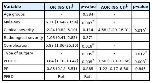
Predictors of the unfavorable outcomes at discharge (N=73)
3. Long-term Postsurgical Outcome
The mean duration of follow-up in our series was 45.1 months (range, 6–92 months). Purely in terms of the stratified CCOS scoring (a score of 11 or more being an indicator of a favorable outcome) [19], all except 1 patient (who died after a second surgery 3 months later) had a favorable outcome (n = 72, 98.6%; mean CCOS score, 13.3; range, 11–16) (Table 1). Almost all of the patients (98.4%) reported a relief of headache and suboccipital pain, out of which, 54.4% patients could completely discontinue their medications. Functionality improved in 59 patients (89.4%), out of which 15.1% patients (n = 10) were completely functional and 74.2% (n = 49) patients could function at > 50% of their preoperative states. With respect to the nonpain symptoms, 86.4% (n = 57) showed improvement, out of which medications could be totally withdrawn in 24.2% patients (n=16) with 13.6% (n = 9) showing no improvement.
On the gestalt questionnaire, 43 patients (58.9%) had an improvement, 27 patients (37%) reported no change while 3 patients deteriorated (4.1%), including one death. Therefore, the comparison of gestalt questionnaire and CCOS scale revealed a pattern similar to the short-term outcome (gestalt method underestimated improvements, 58.9% vs. 98.6%). Many of the patients with so-called unchanged symptoms did, therefore, improve objectively in the long term on CCOS scale. At the same time, we observed that the proportion of delayed deterioration was slightly underestimated by the CCOS score using the score cutoff of 11 (n = 1 [1.4%] vs. n = 3 [4.1%]).
The mean change of CCOS score was 2.15 (median, 2) ranging from -1 to 5. Table 2 shows the scores in various surgical subgroups. However, the proportion of patients with a median positive score change of 2 or more was higher (p = 0.06) in the PFBDD group (77.5%) and least in the PF group (50%). Interestingly, the patient perceived level of long-term satisfaction following surgery correlated well with a positive gain of at least 2 points in the CCOS scoring system (n = 49 [67.1%] satisfied vs. n = 24 [32.9%] dissatisfied, p < 0.001). Three patients (4.1%) developed a delayed clinical deterioration in the follow-up (details shown in Table 3). All the late deteriorations followed PFBDD. Only one patient among the 3 had an assimilated atlas with C2–3 fusion with asymmetric lateral elements. This patient also had a high pB-C2 distance. The average time to clinical deterioration was 4 years. All of them underwent a redo surgery with distraction of the craniocervical junction and surgical fixation.
DISCUSSION
CM-1 is characterized by a narrow posterior fossa leading to the caudal displacement of the tonsils (> 5 mm) and a CSF circulation disruption at the level of the FM [1-4]. Syringomyelia often coexists and symptoms can be myriad. CM-1 may or may not be associated with a bony instability like AAD or BI [1-5,17-18,29-31]. When CM occurs without a bony instability, the condition can be termed as a “pure CM” and traditionally, PFD has remained their treatment of choice [9,26-37]. We found that nearly 31% of the patients had some bony anomaly like basilar impression, platybasia, assimilation of atlas, and C2–3 congenital fusion. However, the incidence of these anomalies did not vary significantly among the 3 treatment groups. Goel [22] analysed the bony anomalies accompanying pure CM and noted that basilar impression or type B BI was present in some of these cases and the anomaly was one of the responsible factors for the reduction in posterior fossa volume. This anomaly, however, does not compress the neural structures and does not represent a bony instability. Eleven of the 40 patients (27.5%) had bony anomaly in the series reported by Salunke et al. [18].
1. Surgical Strategies and Their Efficacies
Surgical decompression remains the mainstay of treatment in symptomatic CM patients. The role of decompressive surgery in asymptomatic or oligosymptomatic CM-1, however, remains controversial. In a recent systematic review, Langridge et al. [23] studied asymptomatic adult CM and found that these patients remain stable despite the presence of syringomyelia and advocated surgical decompression only in overtly and chronically symptomatic patients. Pomeraniec [31], in another study, found that the overwhelming majority of the patients (92.9%) remained clinically stable on a conservative treatment. Nearly 40% of these nonoperated patients even reported an improvement in symptoms. However, their group concluded that symptoms like sleep apnea/dysphagia or the presence of a syrinx must be viewed as surgical indications due to significant improvements found with a timely surgery. In another large series, Strahle7 found that the natural history of conservative management in asymptomatic CM was mainly benign and stable. However, those who show a change in the status, improvements occur less commonly than disease progression. Therefore, there is definitely a role of nonoperative treatment in these patients. That said, in symptomatic patients, presence of large syringomyelia, or setups where patients may not agree for regular follow-up visits, surgical decompression remains a valid choice. Herein comes the importance of choosing a surgical approach with minimum complications.
The majority of our patients underwent a PFD (n = 61, 83.6%). In a meta-analysis, Förander et al. [9], showed that bony decompression only did not differ significantly than the dural decompression with respect to important outcomes parameters like the rates significant postoperative clinical improvement and resolution of syringomyelia. Their study, however, showed that dural decompression could lead to a significant decrease in the reoperation rates related to clinical nonimprovement (2% vs. 8%,) but at the cost of a higher CSF-related complications (7% vs. 0%,) [9]. Lin et al. [10] reported that PFBDD provided a better clinical result in the presence of a syringomyelia. In a recent series on pediatric CM-1, Massimi et al. [38] have found a better result with only bony decompression. Expansile dura allowing an expansion of the space after bony decompression along with a higher risk of CSF leak due to a poorly developed musculature are the primary reasons for preferring only a bony decompression in this age group. Massimi et al. [38], in a meta-analysis, noted that PFBD was sufficient in children without a syringomyelia while PFBDD was the procedure of choice in adults or in large syringomyelia irrespective of age. Our patient cohort had a mix of pediatric and adult patients but 89% of the patients had a syrinx. PFBDD in our experience had a very high share of the complications (80%) and associated significantly with an unfavorable short-term outcome. The higher rate of complications in PFBDD is well known and it evidently negated the advantages of neuraxial decompression in our series. Although the patients undergoing PFBDD were more severe clinically, the same cannot entirely explain the poor short-term outcomes as the PF group also had similar patients albeit with a better outcome. Therefore, associated complication represents the Achilles’ hill for PFBDD. We also noted a delayed deterioration in 3 patients undergoing a PFBDD. This was in contradistinction to the study of Förander et al. [9].
Despite our observations, PFBDD remains the first choice of treatment in adult CM-1, particularly in the presence of a syringomyelia and in resurgery cases [36-38]. Considering the risks of CSF leak that can offset the results of a good surgical decompression, some authors have utilized intraoperative ultrasonography in deciding the need for duraplasty, adding more objectivity in the decision making [39,40]. PFD was surprisingly associated with a comparatively better outcome in this series. The age groups did not differ between the treatment groups. Various authors have found that PFBD was a treatment of choice in children but not in adults. This procedure was done mainly in the minimally symptomatic patients (p = 0.049) and in the short term, the majority of the patients did not report any change in their symptoms (71%). However, a lack of complications characterized PFD group in our series. Förander et al. [9] had noted a higher reoperation rate with PFBD. In our experience, a resurgery was needed in only one patient in this category. To add, these patients had the best follow-up scores amongst the 3 groups.
We also treated some of our patients with a PF, despite the lack of obvious CVJ instability (n = 12). As Table 2 demonstrates, there was no difference amongst the 3 surgical groups with respect to bony anomalies or radiological disease severity. PF in our series was chosen mostly in patients with a clinically severe disease, similar to PFBDD. We saw that this group of patients had the best mean CCOS scores in the postoperative period, primarily from a reduced complication rate. It is interesting to note that 9 of these patients also underwent a removal of the posterior margin of the FM before fixation and distraction. Therefore, a combination of mechanisms might have resulted in a clinical improvement in this group. Moreover, we believe our experience with surgical fixation for bony CVJ anomalies and the volume of cases performed in our centre ensured that complications of fixation were very low. That said, we need to remember that PF has certain inherent issues like an increased cost of treatment, increased hospital stay, possible vascular injury, restricted neck movement, suboccipital hypesthesia, etc. A strategy of uniform C1/2 fixation has been advocated by Goel et al. [11-13] in all CMs. Salunke et al. [17,18] recently examined this strategy where uniform surgical fixation for pure CM-1 was performed and noted that 30% patients did not improve satisfactorily. The authors concluded that a distraction of the odontoid process led to a vertical expansion of the CSF space leading to a symptomatic improvement. Our study has shown that despite a good immediate outcome, PF remains a sparingly used surgical technique for pure CM-1 (16% of the cases) and the long-term benefits are not sustained (mean long term CCOS score being lowest in PF group).
2. Issues With the Outcome Assessment Tools
Traditionally, the outcomes after Chiari decompression have been reported as gestalt improved, unchanged, worse pattern. In 2012, Aliaga et al. [19] proposed a novel scoring system, called the Chicago Chiari Outcome Score (CCOS) to provide an objective way of postoperative outcome assessment. This system has been externally validated as a better outcome assessment tool, despite it not being a traditional preoperative vs postoperative comparison tool. They maintain that the CCOS score captures the preoperative clinical patient status more accurately and hence allows a better outcome assessment than the traditional system. We found a disparity between patients’ perception and CCOS scores in our study [41]. This could be because the CCOS scoring has 4 different components and despite improvements in some of them, a reduction in score in a particular component, felt important by the patient, might have the patient to report an unchanged or a worsened postoperative status. Aliaga et al. [19] noted a good reliability between gestalt outcome and CCOS score but they also noted some outliers where the gestalt outcomes and CCOS scores did not correspond. This was evident in the short-term outcome for PFBDD group. The mean short-term CCOS in this group was worst despite the fact that 47% patients had improved on gestalt system, more than the PFD group (Table 2). This can be explained by the fact that the patients undergoing PFBDD had a significantly higher share of clinically severe patients. Therefore, despite the patient improving subjectively, the scoring did not add upto 11 or more. As far as the long-term outcome was concerned, 98.6% of the patients had a favorable outcome on CCOS scale, whereas the improvement on the gestalt scale was 58.9% (n = 43), indicating that patient perception is often different than the actual changes in the score. Interestingly, a positive change in the score as the duration of follow-up increased correlated well with the patients’ satisfaction (p ≤ 0.001). This leads us to believe that change in score at follow-up, rather than the follow-up score per se, maybe a better matrix of the long-term postoperative outcome assessment. PFBDD group, despite a significantly higher number of unfavorable outcome, showed the maximum mean increase in the follow-up scores. While this finding may be attributed to the low short-term scores, the long-term outcomes of PFBDD are well proven. Another interesting finding of our study was that CCOS score at follow-up revealed a poor outcome in 1 patient only whereas the gestalt outcome score revealed 3 such patients. Therefore, the score can overestimate the improvement as well. This happens in patients with a high CCOS score at discharge who despite a clinical worsening, still manage to have a score of at least ‘11.’ Therefore, the score difference appears to a better matrix of assessing long-term outcomes, both favorable and unfavorable, after surgery in CM-1.
3. Outcome Predictors
Apart from PFBDD, another significant predictor of an unfavorable outcome in our series was the preoperative clinical disease severity. Patients with minimal symptoms or symptoms that are not advanced and do not call for an urgent surgery have always remained a dilemma as far as decompression surgery is concerned [19]. Their decision of surgery is always relative, with a view to stop the disease progression. Our findings suggest that this group of patients does not perceive the benefits of surgery immediately and rather report a worsening. Thus, a proper discussion about the risks and benefits of surgery with the patient and the family members is necessary in these patients. The exact opposite is also true. The importance of preoperative clinical disease severity as a cause of apparent lack of improvement was pointed out by Aliaga et al. [19]. Like minimal symptoms, a patient with an advanced disease also fails to perceive the benefits of surgery. We observed that the difference in the score increased with follow-up, the patient perceived satisfaction also improved significantly. This indicates that minor improvements are picked up only by a validated scoring system and often not reported by the patients. Therefore, the way the outcome is documented is very important and there is a need to use validated outcome scales like the CCOS uniformly, rather than relying entirely on the patient’s response to a gestalt questionnaire [26].
4. Early and Delayed Postoperative Motor Deterioration and Perspectives
We had 4 patients in our series who had a persistent postoperative motor deterioration requiring a second surgical intervention (Table 3). While one such deterioration was detected immediately after surgery, the other 3 occurred after at least 3 years. Immediate postoperative motor deterioration was observed in a total of 4 patients in this series (5.4%), 3 of whom spontaneously improved. Two of these patients had a pre-existing minor motor weakness a couple of them had an associated bony anomaly. None of these patients had a demonstrable preor postoperative instability. Two of them had undergone a PFBDD, 1 patient each had undergone PFBD and PF respectively. Therefore, a pre-existing weakness, acute clival-cervical angulation associated with type B BI and a potential intraoperative cord manipulation (PFBDD in 2 and PF in 1) could be responsible for an immediate postoperative motor deterioration, although we cannot definitely claim so. Therefore, exercising due precaution to avoid excessive manipulation of the area especially in those who already have a motor impairment and utilization of intraoperative monitoring techniques could help prevent this relatively rare but important complication [42].
We encountered 3 late deteriorations in our study (4.1%). In a series of CM-1 patients uniformly treated with PF, Salunke et al. [17,18] noted a delayed deterioration in 3 patients (7.5%). Aliaga et al. [19] noted that clinical deterioration in these patients tends to be apparent only after a year of surgery. All the late motor deteriorations followed PFBDD and after an initial improvement. Imaging before the second surgery did not reveal any iatrogenic instability in these patients but did reveal a persistent syringomyelia or a new onset syringomyelia in all 3 patients with late deteriorations. Therefore, an inadequate PFD with a persistent CSF circulation block and subsequent disease progression was the likely cause for the late deteriorations. Therefore, a late deterioration may merely reflect an inadequate PFD. Other possible causes can be a latent bony instability that is ignored at initial evaluation. Sometimes a significant ventral brainstem compression (pB-C2 distance >9 mm) can also be the cause [15]. Therefore, appropriate case selection for PFD, paying attention to decompress adequately at first surgery, close postoperative follow-up etc. remain possible measures. Although these patients were treated with a PF in our series, the improvement after surgery was only marginal (gain of CCOS score of only 1) while 1 patient died after surgery. Our decision for PF was on a presumptive basis, with an idea of doing the “maximum” i.e., a re-exploration of the PFD and distraction while performing PF, in an effort to provide adequate decompression. In absence of a demonstrable (preoperative radiological and intraoperative inspection of the C1/2 joints) iatrogenic instability in any of these “late deterioration” cases, the salvage PF perhaps benefited by providing a vertical decompression of the FM from the associated distraction. PF as a primary treatment of pure CM-1 may be an overkill [26,42].
5. Limitations of the Study
This study is limited by the retrospective design and a lack of a detailed evaluation of the commonly used radiological metrices. Moreover, the numbers of patients undergoing PF was low and the 3 groups of patients did not have similar number of patients. Therefore, the comparison was not very robust, rendering many observations statistically insignificant. Our study was also limited by a lack of follow-up information at different intervals which could have given a better insight into the long-term outcome dynamics. However, our study attempts to characterize the outcomes of various contemporary surgical strategies employed for pure CM-1 using a validated outcome assessment tool. Our study also introduces certain clinicoradiological and outcome measurement matrices which could be useful in the future research. A randomized trial comparing the various techniques will be the best tool to evaluate the various treatment strategies in CM-1.
CONCLUSION
PFD is the most commonly performed surgery and still remains the gold standard in “pure CM-1.” Current surgical strategies are generally successful in providing a favorable long-term outcome. Outcome measured on CCOS is a composite score and despite a lack of correlation with the gestalt system on a few occasions, should be used to bring in objectivity and uniformity in Chiari research. Minimally symptomatic patients and PFBDD predict a poor short-term postoperative outcome. PFBD appears to be a durable procedure while PFBDD group is marred by complications and late deteriorations. PF does not provide any better results than PFD alone in the long run. Late deteriorations after PFD are rare (4.1%) and represent a continued disease progression from an inadequate primary PFD.
Notes
The authors have nothing to disclose.