Highly Accurate Analysis of the Cervical Neural Tract of the Elderly Using ZOOM DTI
Article information

Abstract
Background/Aims
To investigate the fractional anisotropy (FA) values of the cervical spinal cord in elderly individuals using zonally magnified oblique multislice (ZOOM) diffusion tensor imaging (DTI).
Methods
Fourteen healthy elderly volunteers (group E) and 10 young volunteers (group Y) were enrolled. We assessed the FA, apparent diffusion coefficient (ADC), and λ1–λ3 values using 3-T magnetic resonance imaging. The region of interest was contoured entirely inside the spinal cord, with no gray/white matter distinction, in order to avoid including the cerebrospinal fluid.
Results
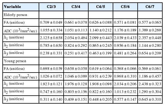
As lower cervical levels were approached, the FA values gradually decreased, while the ADC values increased. The mean FA values at each cervical level were as follows in groups E and Y: 0.71 and 0.70 at the C2/3 level, 0.66 and 0.66 at the C3/4 level, 0.63 and 0.62 at the C4/5 level, 0.57 and 0.57 at the C5/6 level, and 0.58 and 0.57 at the C6/7 level, respectively. The mean ADC values in groups E and Y were 1.06 and 0.99 at the C2/3 level, 1.05 and 1.06 at the C3/4 level, 1.14 and 1.06 at the C4/5 level, 1.18 and 1.21 at the C5/6 level, and 1.39 and 1.46 at the C6/7 level, respectively. There were no significant differences between the elderly and young participants.
Conclusion
In both asymptomatic elderly and young individuals, the FA values gradually decreased and the ADC values increased moving towards lower cervical levels. Age did not affect the FA values, even though mild cord compression was evident due to spondylotic changes. ZOOM DTI has the potential to provide more information than conventional DTI.
INTRODUCTION
Microstructural changes in the spinal cord can result in functional disability. Thus, a method for assessing the area inside and the integrity of a compressed spinal cord is required [1-3]. To date, a number of papers have reported on the use of tractography to depict neuronal fiber tracts. Diffusion tensor imaging (DTI) is widely used to show neuronal fiber tracts around the space occupying lesion in the brain in clinical scene [4]. Some reports have found a relationship between tractography results and prognosis in patients with brain disease. Despite similar trials for spinal cord lesion, conventional DTI could not achieve a high accurate evaluation due to the smaller cross-sectional area of the spinal cord and the variable magnetic susceptibility of the organs surrounding the spinal cord. Magnetic artifacts are often observed during DTI of the cervical spinal cord, because of the unstable magnetic susceptibility of the surrounding regions, such as the lung apex. Moreover, obtaining high resolution images of the cervical spinal cord by DTI prolongs the scan time, which is not desirable in a clinical context; however, lower resolution scans reduce the accuracy of measurements. Given the above, a new technique, termed ZOOM DTI (Philips, Amsterdam, The Netherlands), has been developed [5]. In our previous study, we demonstrated that ZOOM DTI yielded better visibility than did conventional DTI [6]. To estimate the patients with cervical spondylotic myelopathy, we have to clarify the average parameters of neural tracts in elderly person because the majority of patients are advanced age.
In the present study, we measured fractional anisotropy (FA), apparent diffusion coefficient (ADC), and λ1–λ3 values in elderly and young individuals using ZOOM DTI and assessed the differences between these 2 groups. The present study was approved by an Institutional Review Board of Otaru General Hospital (approval number: 30-009) and informed consent was obtained from the participants.
MATERIALS AND METHODS
1. Patient Population
Fourteen asymptomatic healthy elderly persons attending Otaru General Hospital (group E: mean age, 72.9 years; male: female=7:7), and 10 young volunteers (group Y: mean age, 30.2 years; male:female=7:3) were included in the study. Fourteen asymptomatic elderly persons did not have clinical symptoms of acute or slowly progressive spinal cord compressions. However, magnetic resonance imaging (MRI) (e.g., T2-weighted imaging) demonstrated some microstructural changes and slight compression of the cervical spinal cord. Young volunteers were fully informed healthy volunteers without neurologic disease. They were members of our medical imaging department.
2. Imaging Device and Imaging Target
Imaging was performed on both groups using a 3-T MRI scanner (Ingenia, Koninklijke Philips N.V., Amsterdam, The Netherlands).
3. Imaging Parameters
ZOOM DTI imaging sequences were used to obtain cervical spinal DTI images. ZOOM DTI parameters were as follows: repetition time=4,500 msec; echo time=81 msec; field of view (FOV)=72×46 mm2; matrix=70×47; scan time=10:35; single-shot spin-echo; 6MPG direction; slice thickness=5 mm; and 24 slices.
The examination frame ranged from C2 to C7. Diffusion was measured with two b values, 0 mm2/sec. The slab center was placed at C4/5. Volunteers were asked to avoid swallowing during DTI.
Image postprocessing was performed using Fiber Track, the software available on the MRI scanner. The region of interest (ROI) was contoured entirely along the rim of the spinal cord, and was maximally enlarged to include white matter; however, the surrounding cerebrospinal fluid (CSF) was omitted by comparison of the 3-dimensional (3D) ROI square (Fig. 1). The axial plane of each cervical disc level was determined from T2- weighted anatomical images. The parameters analyzed were as follows: FA, ADC, and λ values.
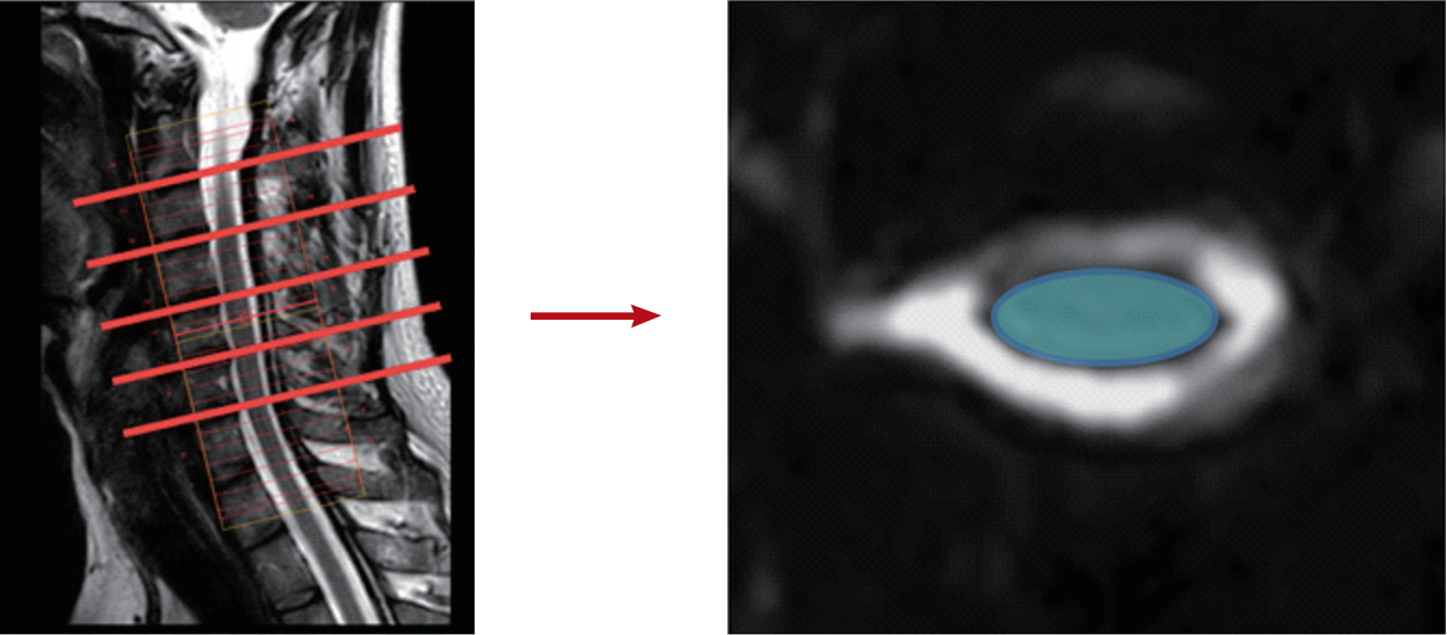
Placement of region of interests in the spinal cord. Contouring the entire cord as wide as possible not to include the surrounding cerebrospinal fluid.
4. Statistical analysis
We used JMP 8 software (SAS Institute Inc., Cary, NC, USA) to perform t-tests for statistical comparison of the data between the 2 groups, with alpha set at 0.05.
RESULTS
All parameters (FA, ADC, and λ1-λ3 values) were successfully acquired in both the E and Y groups (Table 1). There were no significant differences in FA values between groups E and Y. FA values gradually decreased towards lower cervical levels, and there was a significant difference in the FA values between C4/5 and C5/6 (p=0.047).
Comparison of diffusion metrics in the spinal cord of elderly person and young
The mean ADC value was 1.16 for all levels in both groups. There were no significant differences between the groups or between cervical levels. However, ADC values gradually increased toward lower cervical levels.
The mean λ1–λ3 values at all levels were 2.13, 0.91, and 0.42 in group E and 2.06, 0.94, and 0.48 in group Y. There were no significant differences between the 2 groups.
Moreover, the standard deviation and error increased as lower cervical disc levels were approached (Figs. 2, 3). Compared with young individuals, the FA value was slightly higher in asymptomatic elderly individuals, but the difference was not statistically significant.

Diagram of the values of diffusion metrics in cervical cord segments (elderly person). In elderly person group, the standard deviation and error increased as the disc level approached lower. (A) Fractional anisotropy (FA), (B) apparent diffusion coefficient (ADC), (C-E) λ1–λ3.

Diagram of the values of diffusion metrics in cervical cord segments (young person). In young person group, the standard deviation and error increased as the disc level approached lower. This tendency in young person group is same as that in elderly group. (A) Fractional anisotropy (FA), (B) apparent diffusion coefficient (ADC), (C-E) λ1–λ3.
DISCUSSION
In the brain and spinal cord white matter, cellular membranes and myelin sheaths act as natural barriers to molecular diffusion, including the movement of water molecules, which affects isotropy [2].
ZOOM DTI is a recently developed method for the estimation of fiber tracts [5]. Spatial resolution of DTI image is important for highly accurate evaluations. In particular, in small organs, such as the spinal cord, finer resolution that can clearly distinguish the boundary between the target and the surrounding tissue would be preferable. ZOOM DTI has previously been shown to yield better visibility than conventional DTI [6]. In our previous study, we had evaluated the parameters in young healthy volunteers, and analyzed the mean FA values at each disc level, and shown that the values had high inter-rater reliability [6].
Roine et al. [7] have reported that partial volume effect between white matter and CSF could lead to errors parameter estimation. Drawing the ROI contour is generally more difficult at lower cervical levels, even when using ZOOM DTI, due to artifacts caused by air in the lung apex; including subarachnoid CSF space inside the ROI would result in a decreased FA value [3,8], and thus contouring the ROI requires close attention, even with ZOOM DTI. Moreover, the physiological movement of the spinal cord in response to cardiac pulsation and the respiratory cycle could produce artifacts. Cardiac gating, radiofrequency coils, and magnetic resonance signal suppression bands are frequently used to enhance the quality of DTI in the spinal cord [9].
FA and ADC values in elderly individuals were not significantly different from those in young individuals, which implies that the neural fiber tracts were still preserved in an environment of mild spinal degeneration. Most of the patients attending our outpatient clinic are elderly, yet, the results of the present study indicate that it is not necessary to take the age difference into account when assessing DTI parameters. Furthermore, FA values gradually decreased and ADC increased as lower cervical level were approached, which is similar to the findings of a previous report using the conventional DTI method [1]. At the upper spinal level, the area of the central gray matter is smaller compared with the lower level of the spinal cord. Central gray matter of the spinal cord, mostly composed of nuclei, typically shows significantly lower anisotropy than spinal white matter. In this study, ROI included both gray and white matter and resulted in decreased FA values in a caudal direction [10].
The elderly group had a slightly higher FA value than the young group, although the difference was not statistically significantly different. In the elderly group, mild degenerative change with slight compression of the cervical spinal cord, was often observed. Facon et al. [3] reported an acute onset case of extradural spinal metastasis that encased and compressed the thoracic spinal cord, which resulted in a slightly increased FA value as compared to normal control values. Nevertheless, it is noteworthy that, to date, almost all of the previous studies described both decreased FA and increased ADC values for cases of compression of the spinal cords due to cervical spondylosis [8,11-16]. Agosta et al. [17] found a strong negative correlation between normal cervical cord FA and the patient’s age. They associated this finding with the presence of microstructural changes. Mamata et al. [10] found that the ADC value of the normal spine cord showed a positive correlation with age, while the FA value was negatively correlated with age. These results are inconsistent with our study analysis. We suspect that high-resolution assessment achieved by ZOOM DTI caused the difference of results between our study and previous studies. As mentioned earlier, fluctuation in FA and ADC values was strongly influenced by CSF. We could set ROI strictly inside the spinal cord and enlarged maximally, while excluding the possibility of encasement of CSF by checking 3D ROI squares.
Therefore, the relationship between anisotropy and compressed neural fiber tracts should be further investigated using a highly accurate approach, such as ZOOM DTI.
CONCLUSION
ZOOM DTI provides better visibility and higher accuracy for a small FOV than does conventional DTI. FA values gradually decreased and ADC values increased toward lower cervical levels in asymptomatic elderly as well as young individuals, and there was no significant difference between elderly and young despite of mild cervical degenerative change.
Notes
The authors have nothing to disclose.
Acknowledgements
This study was supported by Philips Healthcare.