Warning: mkdir(): Permission denied in /home/virtual/lib/view_data.php on line 81 Warning: fopen(/home/virtual/e-kjs/journal/upload/ip_log/ip_log_2024-04.txt): failed to open stream: No such file or directory in /home/virtual/lib/view_data.php on line 83 Warning: fwrite() expects parameter 1 to be resource, boolean given in /home/virtual/lib/view_data.php on line 84 Introduction of a New Radiographic Parameter to Predict Proximal Junctional Kyphosis in Adult Spinal Deformity: UIVPTA (Uppermost Instrumented Vertebra-Pelvic Tilt Angle)
Neurospine Search

CLOSE


Park, Lee, Park, and Shin: Introduction of a New Radiographic Parameter to Predict Proximal Junctional Kyphosis in Adult Spinal Deformity: UIVPTA (Uppermost Instrumented Vertebra-Pelvic Tilt Angle)

Abstract

Objective

To introduce a new sagittal parameter, uppermost instrumented vertebra-pelvic tilt angle (UIVPTA), and to determine the effects on the proximal junctional kyphosis (PJK) development in adult spinal deformity (ASD) surgery.

Methods

Patients ≥ 60 years with ASD who underwent low thoracic spine to pelvis fusion with a minimum of 2-years of follow-up were included in this study. Two groups were created according to PJK development. Various clinical and radiographic factors were compared between PJK and non-PJK groups to identify the risk factors for PJK. Cutoff value of UIVPTA for PJK development was calculated using receiver operating characteristic curve according to different pelvic incidence groups. Linear regression analysis was performed to identify factors to affect UIVPTA.

Results

One hundred fifity-one patients were included in this study. There were 135 female patients (89.4%). Mean age was 70.5 years. PJK developed in 65 patients (43.0%). Multivariate analysis showed that overcorrection relative to age-adjusted pelvic incidence (PI) minus lumbar lordosis (LL) (PI–LL) target and lower UIVPTA were independent risk factors for PJK. The cutoff value of UIVPTA for PJK development was calculated as 4.0° in patients with PI less than 45°, 9.5° in patients with PI between 45° and 60°, and 13.0° in patients with PI greater than 60°. Linear regression analysis showed that UIVPTA was positively affected by postoperative values of LL (coefficient = 0.505), PI–LL (coefficient = 0.674), and pelvic tilt (coefficient = 0.286).

Conclusion

Optimal correction within the age-adjusted PI–LL combined with keeping UIVPTA within optimal range is suggested for the prevention of PJK.

INTRODUCTION

In surgical treatment for adult spinal deformity (ASD), optimal correction of the sagittal alignment is important for favorable clinical outcomes [1,2]. Since Schwab et al. [3] suggested ideal sagittal alignment goals such as pelvic incidence (PI) minus lumbar lordosis (LL) within±10°, pelvic tilt (PT) ≤ 20°, and sagittal vertical axis (SVA) ≤ 50 mm, these criteria has been conventionally used to determine the optimal sagittal alignment. Although the beneficial effect of these criteria on clinical outcome have been supported in previous studies [4,5], the effect on preventing mechanical failure such as proximal junctional kyphosis (PJK) remains controversial [6-9]. Meanwhile, the concept of age-adjusted sagittal alignment concept was introduced by Lafage et al. [10] suggesting the appropriate sagittal spinopelvic parameters should be assessed in consideration of patient age. Subsequent studies supported this concept, showing that the overcorrection of PI–LL relative to the age-adjusted target increased PJK risk [11-15]. Although the concept has been validated in preventing PJK development, it addresses only the amount of LL.
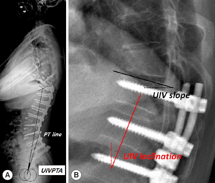
Recently, the clinical significance of UIV orientation such as UIV slope and inclination has been emphasized rather than LL amount itself, given that higher UIV slope and inclination may impose kyphotic force above UIV and subsequently increases PJK development [7,16,17]. Therefore, UIV orientation as well as LL should be considered importantly together in ASD surgery. However, these conventional parameters representing UIV orientation would not be fixed and change according to different standing position. UIV orientation can be affected by pelvic rotation, amount of LL correction, and rod contour above L1. Therefore, UIV orientation needs to be comprehensively assessed with regard to all component from pelvis to UIV. Herein, we introduce a new sagittal parameter, the uppermost instrumented vertebrapelvic tilt angle (UIVPTA), which is non-positional parameter to represent UIV orientation (Fig. 1). The present study primarily aims to demonstrate the clinical significance of UIVPTA with regard to PJK development in patients undergoing long-fusion surgery from lower thoracic vertebra to sacrum for ASD. The secondary aim was to provide the cutoff value of UIVPTA to instigate PJK development.

MATERIALS AND METHODS

This study was approved by the Institutional Review Board of Samsung Medical Center (IRB No. 2022-08-128). The need to obtain informed consent was waived because of the retrospective nature of this study.

1. Study Cohort

This study was a retrospective case series with records retrieved from the prospective ASD database at our institution. Individuals eligible for the study included ASD patients with lumbar degenerative kyphosis (LDK) who underwent surgical correction between 2013 and 2020. The detailed inclusion criteria were as follows: (1) patients with radiographically proven with sagittal malalignment (SVA ≥ 50 mm, PI–LL ≥ 10°, and PT ≥ 25°), (2) patients who showed the cardinal signs of LDK such as stooped gait, inability to lift heavy objects, difficulty in climbing slopes, and need for elbow support in front of sink with evidence of calluses on the extensor surface of the elbow [18,19], (3) patients who underwent long-segment fixation from lower thoracic spine (T9–12) to sacrum or pelvis, (4) patients with previous fusion surgery if the fusion length was ≤ 2 levels, and (5) patients who were followed up more than 2 years.

2. Surgical Techniques

All surgeries were performed by 1 of 3 surgeons in a single center. Corrective surgery was performed either posterior only surgery using posterior column osteotomy with or without pedicle subtraction osteotomy (PSO) or via a combined anteriorposterior approach using oblique or anterior lumbar interbody fusion (ALIF). Although the surgical methods were determined based on the patient’s deformity status, the preferred surgical technique at our institution is the combined anteriorposterior approach. All L5-S1 levels were treated by interbody fusion with either ALIF or posterior lumbar interbody fusion (PLIF). ALIF with hyperlordotic cage was preferred method to maximize the LL, but PLIF was performed in case with unfavorable iliac vessel anatomy, previous abdominal surgery, or severely obese patients. At or above L4–5 levels, lateral approaches using oblique lumbar interbody fusion or anterior column realignment technique were preferred [20]. In case of rigid kyphotic deformity, PSO was performed. Pelvic fixation was routinely performed using conventional iliac screw fixation except for cases with lumbosacral fusion status due to previous fusion surgery or patients with concerns for screw prominence due to shallow soft tissue coverage. All surgeries were performed using an open method with titanium rods. Augmentation techniques at UIV to prevent PJK such as cement augmentation or posterior tether were not used.

3. Definition of PJK

PJK was defined radiographically as a postoperative proximal junctional angle (PJA) ≥ 10° and ≥ 10° greater than the preoperative PJA [21]. However, the current definition of PJK used in the present study broadly included any types of PJK including soft tissue failure, fracture at UIV or UIV+1, fixation failure at implant-bone interface at UIV, and revision surgery. According to PJK development, 2 groups were created: PJK and non-PJK groups. Various clinical parameters were compared between the 2 groups with respect to patient factors, surgical factors, and radiographic parameters.

4. Patient and Surgical Factors

Patient factors included age, sex, T score (gm/cm2) on bone mineral density (BMD), perioperative use of an anabolic agent, body mass index (BMI), American Society of Anesthesiologists (ASA) physical status classification grade, and history of diabetes mellitus (DM). Surgical variables included revision surgery, surgical approach (posterior only surgery or combined anteriorposterior approach), and PSO.

5. Radiographic Evaluation

Radiographic parameters were separately measured with respect to the conventional global parameters and regional parameters. Conventional global parameters included PI, LL, sacral slope (SS), PT, T1 pelvic angle (TPA), and SVA. All parameters were measured on whole-spine 36-inch standing radiographs preoperatively and 2 weeks postoperatively. In addition to comparison of conventional radiographic parameters, 3 categorical criteria were also evaluated. To assess the effect of Schwab suggestion about optimal postoperative PI–LL was classified into 3 groups: > 10°, within±10°, and < -10° [3]. Global alignment and proportion (GAP) scores were calculated and 3 groups were created such as proportioned (score: 0–2), moderately disproportioned (score: 3–6), and severely disproportioned (score ≥ 7) according to the original scoring system [22]. Finally, the effect of the age-adjusted alignment target on PJK development was also analyzed. The age-adjusted PI–LL target was calculated using a previously reported formula: PI–LL= (age–55)/2+3 [10,23]. According to the offset value between the actual and age-adjusted PI–LL values, the patients were divided into 3 groups: undercorrection (offset < -10°), ideal correction (offset within ±10°), and overcorrection (offset > 10°).
Regional parameters included UIVPTA, lower LL (LLL), upper LL (ULL), lumbar distribution index (LDI), UIV-L1 angle, UIV slope, and UIV inclination. UIVPTA was determined by the angle between a line drawn from the center of femoral heads to the UIV center and a line from the center of the femoral head to the midpoint of S1 endplate (PT line) (Fig. 1A). ULL was defined as lordosis between L1 to L4, and LLL was defined as lordosis between L4-S1. LDI was calculated as LLL/LL× 100 (%). UIV-L1 angle was the angle between the cranial endplate of the UIV and the caudal end plate of L1. Positive value of UIV-L1 angle denotes kyphotic curvature. UIV slope is the angle between the UIV superior endplate and a horizontal line (Fig. 1B) [16]. UIV inclination is the angle between the best-fit line crossing the vertebral body center of UIV-2 to UIV and a vertical line (Fig. 1B) [16].
The comparison of radiographic parameters was repeated for patients who achieved the ideal correction relative to the age-adjusted PI–LL target in order to adjust for the effect of age-adjusted PI–LL on UIVPTA.

6. Statistical Analysis

Data are presented as the frequencies with percentages for categorical variables and means with standard deviations for continuous variables. Univariate analyses were performed using the chi-square test or Fisher exact tests for categorical variables, and using independent t-test to assess differences in the continuous variables between the 2 groups. Multivariate logistic regression analysis was performed using all variables that had a significance < 0.05 in univariate analyses to identify the risk factors for PJK development. Cutoff values of UIVPTA for PJK development were calculated using receiver operating characteristic (ROC) curve as the point at which the sensitivity and specificity were equal. In addition, cutoff value of UIVPTA was calculated separately according to PI groups. Linear regression analysis was performed to identify factors to affect UIVPTA. Statistical analyses were conducted by professional statisticians using IBM SPSS Statistics ver. 27.0 (IBM Co., Armonk, NY, USA). A p-value of < 0.05 was considered statistically significant.

RESULTS

Among 452 adult patients who underwent the surgical correction for ASD during the study period, 151 patients met the inclusion criteria and constitute the study cohort; 135 patients (89.4%) were female and mean age was 70.5 years. Mean T score on BMD was -1.6 g/cm2. The combined anteriorposterior approach was performed in 121 patients (80.1%) and PSO in 29 patients (19.2%). During mean follow-up duration of 34.5 months, PJK developed in 65 patients (43.0%). With regard to the types of PJK, there were 30 patients with PJA > 20° without bony failure, 31 patients with fracture at UIV or UIV+1, and 4 patients with screw pullout.
For patient factors, there were no significant differences in terms of age, sex, T score, perioperative use of anabolic agent, BMI, ASA physical status classification grade, and history of DM (Table 1). Surgical variables also did not significantly differ between the 2 groups with respect to revision surgery, surgical approach, and PSO (Table 1).
On univariate analysis of radiographic parameters, conventional global parameters including PI, LL, PI–LL, SS, PT, TPA, and SVA showed no significant differences between the 2 groups. Also, there were no significant differences in patient distribution according to Schwab optimal PI–LL or GAP score between the 2 groups. However, there were significantly more patients with overcorrection relative to the age-adjusted PI–LL target in the PJK group than in the non-PJK group (p= 0.024). In terms of regional parameters, UIVPTA was significantly lower in the PJK group than in the non-PJK group (6.7° vs. 11.1°, p= 0.004) (Table 2). UIV inclination was significantly higher in the PJK group than in the non-PJK group (15.0° vs. 11.3°, p = 0.004) (Table 2).
Multivariate logistic regression analysis demonstrated that overcorrection relative to ideal age-adjusted PI–LL target (odds ratio [OR], 7.274; 95% confidence interval [CI], 1.477–10.752, p= 0.011), UIVPTA (OR, 0.942; 95% CI, 0.987–0.989; p= 0.017), and UIV inclination (OR, 1.066; 95% CI, 1.019–1.115; p= 0.006) were independent risk factors associated with PJK development (Table 3). To eliminate the beneficial effect of undercorrection of PI–LL value on UIVPTA, we repeated univariate analysis of radiographic parameters only for the 90 patients who achieved ideal correction relative to the age-adjusted PI–LL target. In the analysis, only lower UIVPTA was a single significant risk factor for PJK (12.8° in the non-PJK group vs. 7.8° in the PJK group, p= 0.002) (Table 4).
The cutoff value of UIVPTA for PJK development was calculated as 4.0° in patients with PI less than 45°, 9.5° in patients with PI between 45° and 60°, and 13.0° in patients with PI greater than 60° (Table 5). Linear regression analysis showed that UIVPTA was significantly affected by postoperative values of LL (coefficient= 0.505), PI–LL (coefficient= 0.674), and PT (coefficient= 0.286) (Table 6).

1. Representative Cases

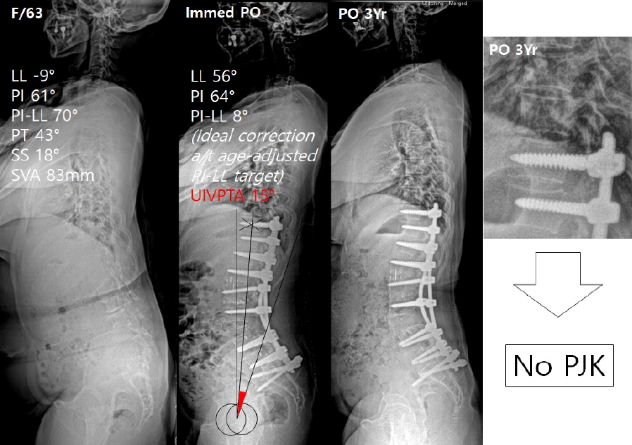
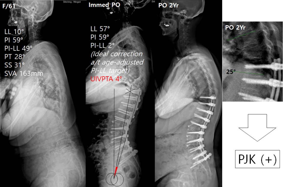
Two representative cases are presented in Figs. 2 and 3. In both cases, the PI–LL was corrected within the ideal range relative to age-adjusted PI–LL target. However, UIVPTA was smaller (4°) in patient of Fig. 3 than in patient of Fig. 2 (15°). At the last follow-up, PJK occurred in patient of Fig. 2.

DISCUSSION

LL correction is a key step for obtaining optimal sagittal alignment in ASD surgery. There have been several guidelines such as Schwab PI–LL criteria or Lafage age-considered sagittal alignment concept, regarding the degree of LL that should be corrected [10,24]. Although these criteria have been validated for preventing mechanical failure such as PJK [13,25], several aspects were still overlooked such as the shape of LL (represented by LDI) and contour of the construct above L1, particularly in cases of proximal fixation extending beyond L1. UIV would be differently positioned even under the same amount of LL. Recent studies demonstrated that the dorsal orientation of the UIV, by imposing the reciprocal kyphotic forces above the UIV, is more important than LL itself as a risk factor for PJK development [26-29]. Therefore, UIV orientation and degree of LL should be both important considerations. This fact inspired us to develop a new radiographic parameter, UIVPTA, which reflects both the degree of LL and UIV orientation.
The clinical utility of UIVPTA in predicting PJK development was demonstrated in this study. We showed that lower UIVPTA was a significant risk factor for PJK in univariate and multivariate analyses. Lower value of UIVPTA equates the UIV movement away from the vertical axis and toward the PT line thereby potentially increasing UIV slope or inclination. However, lower UIVPTA does not necessarily means higher UIV slope or UIV inclination because UIVPTA include the pelvis position and UIV slope or inclination does not. Assuming the pelvis is fixed, the change of UIVPTA may be directly reflected on the changes of UIV slope or inclination. However, because the pelvis is not fixed structure, UIVPTA can be also affected by pelvic position. UIVPTA would decrease by anterior rotation of pelvis and conversely increase by posterior pelvic rotation. We could calculate the cutoff value of UIVPTA using ROC analysis. The cutoff values were differently presented according to PI. We can expect that patients with low PI would have smaller UIVPTA and patients with high PI have higher UIVPTA. As to our expectation, the ROC analysis showed that the cutoff values were smallest in low PI patients and greatest in high PI patients.
UIVPTA is an angle which is formed by the influence of several parameters. To identify the parameters to affect UIVPTA, linear regression analysis was performed. It reveals that UIVPTA was positively affected by LL, PI–LL, and PT. This result indicates that UIVPTA is not simply explained by conventional over- or undercorrection of LL. With regard to correction amount, greater LL means overcorrection of LL, while greater PI–LL and PT means undercorrection. In case of overcorrection, greater LL tends to shift UIV posteriorly (toward increasing UIVPTA) and rotate pelvis anteriorly (toward decreasing UIVPTA) at the same time. Therefore, simply correction amount of LL is not enough to predicting UIV orientation. We thought that the issue of correction amount needs to be assessed including pelvic rotation. In that sense, UIVPTA could be used to appropriately define the over- and undercorrection.
In the linear regression analysis, UIVPTA was also positively affected by PI–LL. This indicates that high PI–LL, which means undercorrection, leads to greater UIVPTA and decreases the PJK risk. However, based on the previous reports, undercorrection should be avoided due to an association with worse clinical outcomes [6,30,31]. Therefore, undercorrection beyond the designated degree cannot be permitted just to increase UIVPTA. Considering the clinical importance of the age-adjusted PI–LL target on PJK development in the previous studies and the current study, we assumed that the maximum degree of postoperative PI–LL gap should be within the range of the age-adjusted PI–LL target. Therefore, we surmised the necessity of repeating an analysis with eliminating the potential beneficial effect of undercorrection on UIVPTA by including only patients who achieved ideal correction. Re-analysis also demonstrated that only UIVPTA was a significant risk factor for PJK development. This result suggests that UIVPTA combined with age-adjusted PI–LL target can better predict PJK development compared with use of only age-adjusted PI–LL target.
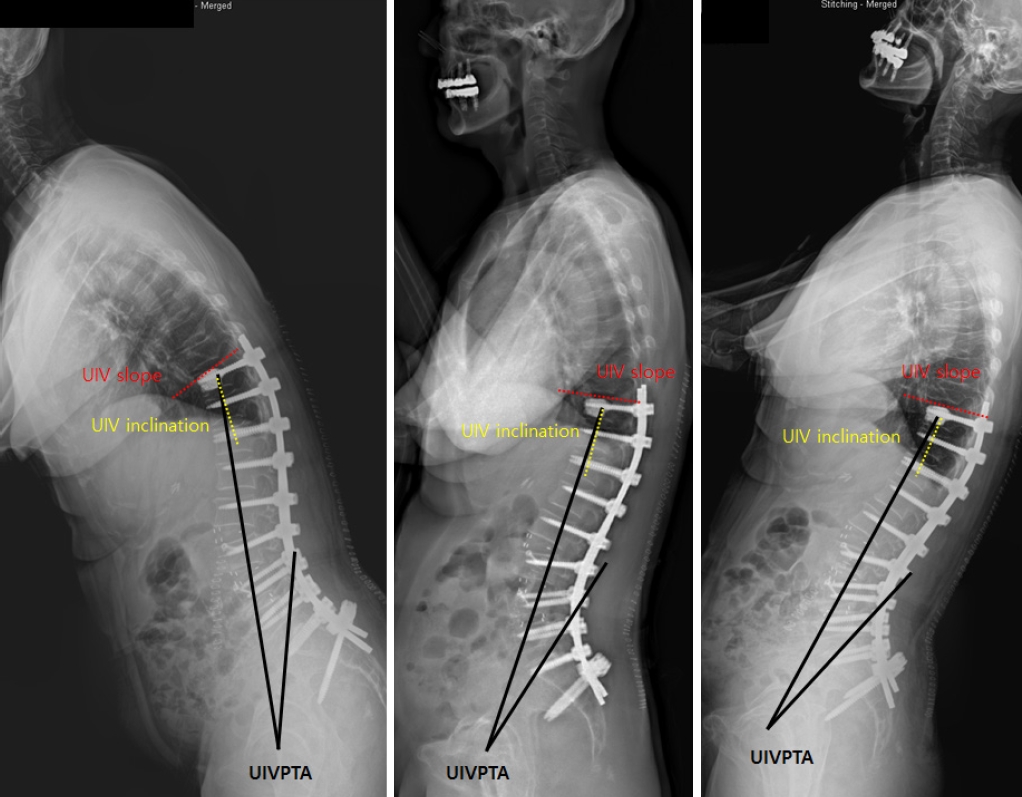
Lastly, the UIVPTA has additional advantages regarding the measurement issue. Because UIVPTA is non-positional parameter, the postoperative UIVPTA will not change according to patient’s position (Fig. 4). Our concept of UIVPTA is similar to the background of introduction of TPA, which is irrelevant to the posture unlike PT in 2014 [32]. However, unlike TPA, UIVPTA can be measured during surgery and it would not change postoperatively, although all UVPTAs were measured postoperatively in this study. This study has a few limitations. First, this study was performed with patients having UIV at lower thoracic spine (T9–12). Therefore, it may be applied universally to patients with UIV of mid- and upper thoracic spine. A single UIVPTA value may not be applied when UIV is located at mid-to upper thoracic vertebra because the kyphotic curvature of thoracic spine. However, it is reported that the mode of PJK is different between upper and lower thoracic vertebra as site of UIV [33,34]. The incidence of PJK is reported to be higher in UIV at lower thoracic spine compared to upper thoracic spine. With regard to the failure mode, the fracture type PJK develops more frequently in patients with UIV at lower thoracic vertebra and PJK with soft tissue failure or spondylolisthesis occurs more in patients with UIV at upper thoracic vertebra. In addition, there are few patients who require long fusion to upper thoracic vertebra due to LDK in our institution although LDK is the leading diagnoses which requires a long fusion surgery. Because the main deformed pathologic lesion in LDK is located within lumbar spine, it is common to stop at the lower thoracic spine. These are the reason why we included only patients with UIV at lower thoracic vertebra. Second, we defined PJK as any form of kyphosis with PJA > 20°. Therefore, the PJK in this study indicates the radiographic term without clinical consideration. Thus, PJK group might include the patients without significant clinical deterioration. However, a recent study demonstrated that even if soft tissue type PJK was asymptomatic at initial development, it progressed radiographically with time and eventually gave a negative impact on the clinical outcomes in longterm follow-up [35]. In addition, PJK group in this study included patients with proximal junctional failure (PJF) such as soft tissue failure, fracture or screw pullout. It is well known that the clinical outcome was significantly inferior in patients with PJF compared to those without PJF [36,37]. Therefore, we believe that all radiographic PJKs should be considered importantly regardless of the initial symptom. Despite the limitations, we believe that UIVPTA could provide a new guide for PJK prevention. More importantly, UIVPTA can be measured intraoperatively and does not change with position.

CONCLUSION

Overcorrection relative to the age-adjusted PI–LL target and lower UIVPTA were independent risk factors for PJK. Therefore, optimal correction within the age-adjusted PI–LL combined with keeping UIVPTA within optimal range is suggested for the prevention of PJK.

NOTES

Conflict of Interest

The authors have nothing to disclose.

Funding/Support

This study received no specific grant from any funding agency in the public, commercial, or not-for-profit sectors.

Author Contribution

Conceptualization: SJP, CSL; Data curation: SJP, JSP, TSS; Formal analysis: SJP, JSP; Methodology: TSS; Project administration: SJP; Writing - original draft: SJP; Writing - review & editing: SJP.

Fig. 1.
(A) Lateral standing radiograph showing UIVPTA. (B) Lateral standing radiograph showing UIV slope and UIV inclination. UIV, uppermost instrumented vertebra; PT, pelvic tilt; UIVPTA, uppermost instrumented vertebra.
ns-2346420-210f1.jpg
Fig. 2.
A 63-year-old-woman presented with severe kyphotic deformity in lumbar spine with PI–LL of 70°. After surgery including 3-column osteotomy and multilevel oblique lumbar interbody fusion, LL was corrected to 56° with PI–LL of 8°, which belongs within age-adjusted ideal correction target. UIVPTA was measured 15° postoperatively. During 3-year follow-up, proximal junctional kyphosis (PJK) did not develop. PI, pelvic incidence; LL, lumbar lordosis; PT, pelvic tilt; SS, sacral slope; SVA, sagittal vertical axis; UIVPTA, uppermost instrumented vertebra-pelvic tilt angle; Immed PO, immediate postoperatively.
ns-2346420-210f2.jpg
Fig. 3.
A 61-year-old-woman with lumbar degenerative kyphosis underwent surgical correction using multilevel anterior column realignments. After surgery, LL was corrected from 10° to 57° and postoperative PI–LL was 2°. Although postoperative PI– LL belongs within age-adjusted ideal correction target, the value of UIVPTA was relatively small (4°). At 2 years postoperatively, proximal junctional kyphosis (PJK) developed along with fractures at UIV and UIV+1. PI, pelvic incidence; LL, lumbar lordosis; PT, pelvic tilt; SS, sacral slope; SVA, sagittal vertical axis; UIVPTA, uppermost instrumented vertebra-pelvic tilt angle; Immed PO, immediate postoperatively.
ns-2346420-210f3.jpg
Fig. 4.
Lateral standing radiographs showing the changes of UIV slope, UIV inclination, and UIVPTA according to different standing position. Note that UIV slope and inclination are changed according to different standing position, but UIVPTA is fixed regardless of patient’s position. UIV, uppermost instrumented vertebra; UIVPTA, uppermost instrumented vertebra-pelvic tilt angle.
ns-2346420-210f4.jpg
Table 1.
Univariate analysis of patient and surgical factors for PJK development
Variable Non-PJK (N = 86) PJK (N = 65) p-value
Age (yr) 69.6 ± 6.6 69.1 ± 7.1 0.677
Female sex 74 (86.0) 61 (93.8) 0.182
T score (g/cm2) on BMD -1.3 ± 1.5 -1.5 ± 1.2 0.452
Perioperative use of anabolic agent 18 (20.9) 14 (21.5) 0.716
BMI (kg/m2) 26.2 ± 3.2 25.4 ± 3.9 0.173
ASA PS classification grade 2.2 ± 0.4 2.1 ± 0.4 0.061
Diabetes mellitus 16 (18.6) 10 (15.4) 0.668
Revision surgery 34 (39.5) 24 (36.9) 0.866
Combined anteriorposterior approach 69 (80.2) 52 (80.0) 1.000
PSO 13 (15.1) 16 (24.6) 0.151

Values are presented as the mean±standard deviation or as number (%).

PJK, proximal junctional kyphosis; BMD, bone mineral density; BMI, body mass index; ASA PS, American Society of Anesthesiologists physical status; PSO, pedicle subtraction osteotomy.

Table 2.
Univariate analysis of radiographic parameters for PJK development in all patients
Variable Non-PJK (N = 86) PJK (N = 65) p-value
Conventional global parameters
Preoperative PI (°) 53.5 ± 11.2 52.5 ± 10.5 0.579
Preoperative LL (°) 10.9 ± 22.7 11.1 ± 19.9 0.950
Preoperative PI–LL (°) 42.6 ± 21.3 41.4 ± 19.2 0.717
Preoperative SS (°) 18.3 ± 11.4 20.6 ± 12.1 0.240
Preoperative PT (°) 34.6 ± 11.4 32.9 ± 11.7 0.360
Preoperative TPA (°) 35.9 ± 14.5 33.8 ± 13.3 0.561
Preoperative SVA (mm) 14.9 ± 38.7 17.8 ± 32.4 0.617
Change in LL (°) 37.2 ± 23.1 39.8 ± 19.7 0.463
Change in PT (°) 17.5 ± 10.7 15.5 ± 12.1 0.287
Change in TPA (°) 22.2 ± 16.0 22.7 ± 12.5 0.585
Change in SVA (mm) 71.4 ± 62.5 54.7 ± 66.7 0.121
Postoperative LL (°) 48.1 ± 12.7 51.0 ± 12.1 0.166
Postoperative PI–LL (°) 5.6 ± 10.3 1.8 ± 9.6 0.022*
Postoperative SS (°) 35.4 ± 9.1 36.2 ± 9.5 0.621
Postoperative PT (°) 17.1 ± 7.3 17.4 ± 8.3 0.830
Postoperative TPA (°) 14.3 ± 8.5 13.5 ± 7.8 0.585
Postoperative SVA (mm) 14.9 ± 38.7 17.8 ± 32.4 0.617
Categories by Schwab optimal PI–LL range 0.243
PI–LL > 10° 26 (30.2) 12 (18.5)
PI–LL within ± 10° 55 (64.0) 48 (73.8)
PI–LL < -10° 5 (5.8) 5 (7.7)
Categories by GAP score 0.321
Proportioned 23 (35.4) 21 (24.4)
Moderately disproportioned 31 (47.7) 46 (53.5)
Severely disproportioned 11 (16.9) 19 (22.1)
Categories by age-adjusted ideal PI–LL target 0.026*
Undercorrection 8 (9.3) 2 (3.1)
Ideal correction 56 (65.1) 34 (52.3)
Overcorrection 22 (25.6) 29 (44.6)
Regional parameter
UIVPTA (°) 11.1 ± 7.2 6.7 ± 6.8 0.004*
LLL (°) 29.0 ± 10.6 31.3 ± 9.5 0.168
ULL (°) 19.1 ± 11.8 19.7 ± 11.0 0.773
LDI (%) 62.4 ± 17.3 61.5 ± 20.0 0.774
UIV-L1 angle (°) 11.1 ± 7.3 10.7 ± 6.8 0.548
UIV slope (°) 5.2 ± 7.3 5.6 ± 8.4 0.759
UIV inclination (°) 11.3 ± 6.9 15.0 ± 8.9 0.004*

Values are presented as the mean±standard deviation or as number (%).

PJK, proximal junctional kyphosis; PI, pelvic incidence; LL, lumbar lordosis; SS, sacral slope; PT, pelvic tilt; TPA, T1 pelvic angle; SVA, sagittal vertical axis; GAP, global alignment and proportion; UIV, uppermost instrumented vertebra; UIVPTA, uppermost instrumented vertebra-pelvic tilt angle; LLL, lower lumbar lordosis; ULL, upper lumbar lordosis; LDI, lumbar distribution index.

* p<0.05, statistically significant difference.

Age-adjusted ideal PI–LL target was calculated as follows: age-adjusted ideal PI–LL=(age–55)/2+3.

Undercorrection means offset value between the actual and age-adjusted ideal PI–LL < -10°, ideal correction means offset value within ±10°, and overcorrection means offset value >10°.

Table 3.
Multivariate analysis of risk factors for PJK development
Variable Odds ratio (95% CI) p-value
Categories by age-adjusted ideal PI–LL target
 Undercorrection Reference
 Ideal correction 3.217 (0.458–14.478) 0.285
 Overcorrection 7.274 (1.477–10.752) 0.011*
 UIVPTA (°) 0.942 (0.897–0.989) 0.017*
 UIV inclination (°) 1.066 (1.019–1.115) 0.006*

PJK, proximal junctional kyphosis; CI, confidence interval; PI, pelvic incidence; LL, lumbar lordosis; UIV, uppermost instrumented vertebra; UIVPTA, uppermost instrumented vertebra-pelvic tilt angle.

* p<0.05, statistically significant difference.

Age-adjusted ideal PI–LL target was calculated as follows: age-adjusted ideal PI–LL=(age–55)/2+3. Undercorrection means offset value between the actual and ageadjusted ideal PI–LL < -10°, ideal correction means offset value within ±10°, and overcorrection means offset value >10°.

Table 4.
Univariate analysis of radiographic parameters for PJK development in patients who achieved ideal correction of ageadjusted PI–LL alignment (N=90)
Variable Non-PJK (N = 56) PJK (N = 34) p-value
Conventional global parameter
Preoperative PI (°) 55.1 ± 9.9 53.1 ± 11.1 0.366
Preoperative LL (°) 12.2 ± 20.9 7.4 ± 19.1 0.277
Preoperative PI–LL (°) 42.9 ± 19.9 45.7 ± 19.6 0.523
Preoperative SS (°) 20.7 ± 11.1 17.6 ± 11.9 0.209
Preoperative PT (°) 34.4 ± 11.0 35.2 ± 12.2 0.757
Preoperative TPA (°) 36.5 ± 13.6 38.8 ± 15.7 0.738
Preoperative SVA (mm) 66.5 ± 60.3 89.4 ± 70.9 0.108
Change in LL (°) 34.9 ± 21.1 39.6 ± 21.2 0.306
Change in PT (°) 15.1 ± 11.9 16.3 ± 11.7 0.637
Change in TPA (°) 20.6 ± 16.1 23.8 ± 15.9 0.570
Change in SVA (mm) 46.7 ± 62.4 70.5 ± 75.4 0.109
Postoperative LL (°) 47.0 ± 9.3 46.9 ± 11.3 0.972
Postoperative PI–LL (°) 8.3 ± 5.7 6.4 ± 5.8 0.125
Postoperative SS (°) 35.8 ± 8.3 34.3 ± 10.0 0.456
Postoperative PT (°) 19.4 ± 6.4 18.9 ± 7.4 0.764
Postoperative TPA (°) 15.9 ± 6.9 15.0 ± 7.4 0.586
Postoperative SVA (mm) 19.9 ± 30.9 18.9 ± 40.4 0.895
Categories by conventional optimal PI–LL range 0.819
PI–LL > 10° 18 (32.1) 10 (29.4)
-10° ≤ PI–LL ≤ 10° 38 (67.9) 24 (70.6)
PI–LL < -10° - -
Categories by GAP score 0.896
Proportioned 13 (23.2) 8 (23.5)
Moderately disproportioned 32 (57.1) 18 (52.9)
Severely disproportioned 11 (19.6) 8 (23.5)
Regional parameter
UIVPTA (°) 12.8 ± 5.7 7.8 ± 5.7 0.002*
LLL (°) 28.7 ± 9.3 29.6 ± 9.4 0.645
ULL (°) 18.3 ± 11.0 17.3 ± 9.6 0.657
LDI (%) 62.2 ± 20.5 63.6 ± 18.1 0.752
UIV-L1 angle (°) 11.5 ± 7.1 12.3 ± 6.9 0.091
UIV slope (°) 4.6 ± 7.7 2.8 ± 7.2 0.296
UIV inclination (°) 10.8 ± 6.7 11.9 ± 8.1 0.501

Values are presented as the mean±standard deviation or as number (%).

PJK, proximal junctional kyphosis; PI, pelvic incidence; LL, lumbar lordosis; SS, sacral slope; PT, pelvic tilt; TPA, T1 pelvic angle; SVA, sagittal vertical axis; GAP, global alignment and proportion; UIV, uppermost instrumented vertebra; UIVPTA, uppermost instrumented vertebra-pelvic tilt angle; LLL, lower lumbar lordosis; ULL, upper lumbar lordosis; LDI, lumbar distribution index.

* p<0.05, statistically significant difference.

Table 5.
Cutoff value of UIVPTA to develop PJK according to pelvic incidence
Variable Cutoff value AUC (95% CI) Sensitivity Specificity p-value
PI < 45° (N = 35) 4.0° 0.618 (0.462–0.773) 0.622 0.633 0.004*
45° ≤ PI < 60° (N = 74) 9.5° 0.712 (0.603–0.820) 0.658 0.627 < 0.001*
PI ≥ 60° (N = 42) 13.0° 0.669 (0.520–0.818) 0.694 0.746 0.011*

UIV, uppermost instrumented vertebra; UIVPTA, uppermost instrumented vertebra-pelvic tilt angle; PJK, proximal junctional kyphosis; AUC, area under the curve; CI, confidence interval; PI, pelvic incidence.

* p<0.05, statistically significant difference.

Table 6.
Linear regression analysis showing factors to affect UIVPTA
Variable Unstandardized B Coefficients SE Standardized coefficients beta t p-value
(Constant) 7.661 2.653 2.887 0.005
PI -0.286 0.162 -0.436 -1.772 0.079
LL 0.505 0.197 0.857 2.563 0.012*
PI–LL 0.674 0.175 0.928 3.845 < 0.001*
PT 0.286 0.074 0.328 3.880 < 0.001*
LDI 0.060 0.090 0.165 0.662 0.077
UIV-L1 angle -0.027 0.058 -0.029 -0.475 0.636

UIV, uppermost instrumented vertebra; PT, pelvic tilt; UIVPTA, uppermost instrumented vertebra-pelvic tilt angle; SE, standard error; PI, pelvic incidence; LL, lumbar lordosis; LDI, lumbar distribution index.

* p<0.05, statistically significant difference.

REFERENCES

1. Glassman SD, Berven S, Bridwell K, et al. Correlation of radiographic parameters and clinical symptoms in adult scoliosis. Spine (Phila Pa 1976) 2005;30:682-8.
crossref pmid
2. Kim HJ, Yang JH, Chang DG, et al. Adult spinal deformity: a comprehensive review of current advances and future directions. Asian Spine J 2022;16:776-88.
crossref pmid pmc pdf
3. Schwab F, Patel A, Ungar B, et al. Adult spinal deformity-postoperative standing imbalance: how much can you tolerate? An overview of key parameters in assessing alignment and planning corrective surgery. Spine (Phila Pa 1976) 2010;35:2224-31.
pmid
4. Bess S, Schwab F, Lafage V, et al. Classifications for adult spinal deformity and use of the Scoliosis Research Society-Schwab Adult Spinal Deformity Classification. Neurosurg Clin N Am 2013;24:185-93.
crossref pmid
5. Terran J, Schwab F, Shaffrey CI, et al. The SRS-Schwab adult spinal deformity classification: assessment and clinical correlations based on a prospective operative and nonoperative cohort. Neurosurgery 2013;73:559-68.
pmid
6. Lee CS, Park JS, Nam Y, et al. Long-term benefits of appropriately corrected sagittal alignment in reconstructive surgery for adult spinal deformity: evaluation of clinical outcomes and mechanical failures. J Neurosurg Spine 2020;34:390-8.
crossref pmid
7. Lee JK, Hyun SJ, Kim KJ. Reciprocal changes in the wholebody following realignment surgery in adult spinal deformity. Asian Spine J 2022;16:958-67.
crossref pmid pmc pdf
8. Park SJ, Lee CS, Chung SS, et al. Different risk factors of proximal junctional kyphosis and proximal junctional failure following long instrumented fusion to the sacrum for adult spinal deformity: survivorship analysis of 160 patients. Neurosurgery 2017;80:279-86.
crossref pmid pdf
9. Park SJ, Lee CS, Park JS, et al. Should thoracolumbar junction be always avoided as upper instrumented vertebra in long instrumented fusion for adult spinal deformity?: risk factor analysis for proximal junctional failure. Spine (Phila Pa 1976) 2020;45:686-93.
pmid
10. Lafage R, Schwab F, Challier V, et al. Defining spino-pelvic alignment thresholds: should operative goals in adult spinal deformity surgery account for age? Spine (Phila Pa 1976) 2016;41:62-8.
pmid
11. Byun CW, Cho JH, Lee CS, et al. Effect of overcorrection on proximal junctional kyphosis in adult spinal deformity: analysis by age-adjusted ideal sagittal alignment. Spine J 2022;22:635-45.
crossref pmid
12. Kim HJ, Yang JH, Chang DG, et al. Proximal junctional kyphosis in adult spinal deformity: definition, classification, risk factors, and prevention strategies. Asian Spine J 2022;16:440-50.
crossref pmid pdf
13. Lafage R, Schwab F, Glassman S, et al. Age-adjusted alignment goals have the potential to reduce PJK. Spine (Phila Pa 1976) 2017;42:1275-82.
crossref pmid
14. Park SJ, Lee CS, Kang BJ, et al. Validation of age-adjusted ideal sagittal alignment in terms of proximal junctional failure and clinical outcomes in adult spinal deformity. Spine (Phila Pa 1976) 2022;47:1737-45.
crossref pmid
15. Rodnoi P, Le H, Hiatt L, et al. Ligament augmentation with mersilene tape reduces the rates of proximal junctional kyphosis and failure in adult spinal deformity. Neurospine 2021;18:580-6.
crossref pmid pmc pdf
16. Lafage R, Line BG, Gupta S, et al. Orientation of the uppermost instrumented segment influences proximal junctional disease following adult spinal deformity surgery. Spine (Phila Pa 1976) 2017;42:1570-7.
crossref pmid
17. Moon BJ, Han MS, Kim JY, et al. Thoracolumbar slope is useful parameter for evaluating health-related quality of life and sagittal imbalance aggravation in adult spinal deformity: a prospective observational cohort study. Neurospine 2021;18:467-74.
crossref pmid pmc pdf
18. Lee CS, Lee CK, Kim YT, et al. Dynamic sagittal imbalance of the spine in degenerative flat back: significance of pelvic tilt in surgical treatment. Spine (Phila Pa 1976) 2001;26:2029-35.
pmid
19. Takemitsu Y, Harada Y, Iwahara T, et al. Lumbar degenerative kyphosis. Clinical, radiological and epidemiological studies. Spine (Phila Pa 1976) 1988;13:1317-26.
pmid
20. Park JS, Lee CS, Choi YT, et al. Usefulness of anterior column release for segmental lordosis restoration in degenerative lumbar kyphosis. J Neurosurg Spine 2021;36:422-8.
crossref pmid
21. Glattes RC, Bridwell KH, Lenke LG, et al. Proximal junctional kyphosis in adult spinal deformity following long instrumented posterior spinal fusion: incidence, outcomes, and risk factor analysis. Spine (Phila Pa 1976) 2005;30:1643-9.
pmid
22. Yilgor C, Sogunmez N, Boissiere L, et al. Global alignment and proportion (GAP) score: development and validation of a new method of analyzing spinopelvic alignment to predict mechanical complications after adult spinal deformity surgery. J Bone Joint Surg Am 2017;99:1661-72.
crossref pmid
23. Passias PG, Jalai CM, Diebo BG, et al. Full-body radiographic analysis of postoperative deviations from age-adjusted alignment goals in adult spinal deformity correction and related compensatory recruitment. Int J Spine Surg 2019;13:205-14.
crossref pmid pmc
24. Schwab F, Ungar B, Blondel B, et al. Scoliosis Research Society-Schwab adult spinal deformity classification: a validation study. Spine (Phila Pa 1976) 2012;37:1077-82.
pmid
25. Jacobs E, van Royen BJ, van Kuijk SMJ, et al. Prediction of mechanical complications in adult spinal deformity surgery-the GAP score versus the Schwab classification. Spine J 2019;19:781-8.
crossref pmid
26. Moon HJ, Bridwell KH, Theologis AA, et al. Thoracolumbar junction orientation: a novel guide for sagittal correction and proximal junctional kyphosis prediction in adult spinal deformity patients. Neurosurgery 2020;88:55-62.
crossref pmid pdf
27. Wu HH, Chou D, Hindoyan K, et al. Upper instrumented vertebra-femoral angle and correlation with proximal junctional kyphosis in adult spinal deformity. Spine Deform 2022;10:449-55.
crossref pmid pdf
28. Xi Z, Duan PG, Mummaneni PV, et al. Posterior displacement of L1 may be a risk factor for proximal junctional kyphosis after adult spinal deformity correction. Global Spine J 2023;13:1042-8.
crossref pmid pdf
29. Xu F, Sun Z, Li W, et al. Correlation between lordosis distribution index, lordosis tilt, and occurrence of proximal junctional kyphosis following surgery for adult degenerative scoliosis. Eur Spine J 2022;31:267-74.
crossref pmid pdf
30. Scheer JK, Lafage R, Schwab FJ, et al. Under correction of sagittal deformities based on age-adjusted alignment thresholds leads to worse health-related quality of life whereas over correction provides no additional benefit. Spine (Phila Pa 1976) 2018;43:388-93.
crossref pmid
31. Yamato Y, Hasegawa T, Togawa D, et al. Rigorous correction of sagittal vertical axis is correlated with better ODI outcomes after extensive corrective fusion in elderly or extremely elderly patients with spinal deformity. Spine Deform 2019;7:610-8.
crossref pmid
32. Pahys JM. T1 pelvic angle (TPA): another acronym to add to the pile, or the missing link for assessing sagittal plane alignment in adult spinal deformity? J Bone Joint Surg Am 2014;96:e172.
crossref pmid
33. Fujimori T, Inoue S, Le H, et al. Long fusion from sacrum to thoracic spine for adult spinal deformity with sagittal imbalance: upper versus lower thoracic spine as site of upper instrumented vertebra. Neurosurg Focus 2014;36:E9.
crossref
34. Yagi M, Rahm M, Gaines R, et al. Characterization and surgical outcomes of proximal junctional failure in surgically treated patients with adult spinal deformity. Spine (Phila Pa 1976) 2014;39:E607-14.
crossref pmid
35. Park SJ, Park JS, Nam YJ, et al. The long-term fate of asymptomatic proximal junctional kyphosis following long instrumented fusion in elderly patients with sagittal imbalance. Spine (Phila Pa 1976) 2021;46:E1097-104.
crossref pmid
36. Park SJ, Park JS, Nam Y, et al. Who will require revision surgery among neurologically intact patients with proximal junctional failure after surgical correction of adult spinal deformity? Spine (Phila Pa 1976) 2021;46:520-9.
crossref pmid
37. Raj A, Lee CS, Park JS, et al. Characteristics of patients undergoing revision surgery for proximal junctional failure after adult spinal deformity surgery: revalidation of the Hart-International Spine Study Group proximal junctional kyphosis severity scale. J Neurosurg Spine 2022 Mar 25:1-8. doi: 10.3171/2022.2.SPINE211387. [Epub].
crossref


Editorial Office
CHA University, CHA School of Medicine Bundang Medical Center
59 Yatap-ro, Bundang-gu, Seongnam-si, Gyeonggi-do 13496, Korea
Tel: +82-31-780-1924  Fax: +82-31-780-5269  E-mail: support@e-neurospine.org
The Korean Spinal Neurosurgery Society
#407, Dong-A Villate 2nd Town, 350 Seocho-daero, Seocho-gu, Seoul 06631, Korea
Tel: +82-2-585-5455  Fax: +82-2-2-523-6812  E-mail: ksns1987@gmail.com
Business License No.: 209-82-62443

Copyright © The Korean Spinal Neurosurgery Society.

Developed in M2PI

Close layer
prev next