Warning: mkdir(): Permission denied in /home/virtual/lib/view_data.php on line 81 Warning: fopen(/home/virtual/e-kjs/journal/upload/ip_log/ip_log_2024-04.txt): failed to open stream: No such file or directory in /home/virtual/lib/view_data.php on line 83 Warning: fwrite() expects parameter 1 to be resource, boolean given in /home/virtual/lib/view_data.php on line 84 The Posterior Cranial Vertical Line: A Novel Radiographic Marker for Classifying Global Sagittal Alignment
Neurospine Search

CLOSE


Park, Hassan, Ferrer, Morrissette, Lee, Cerpa, Sardar, Kelly, Bourret, Hasegawa, Wong, Liu, Hey, Riahi, Huec, and Lenke: The Posterior Cranial Vertical Line: A Novel Radiographic Marker for Classifying Global Sagittal Alignment

Abstract

Objective

To define a novel radiographic measurement, the posterior cranial vertical line (PCVL), in an asymptomatic adult population to better understand global sagittal alignment.

Methods

We performed a multicenter retrospective review of prospectively collected radiographic data on asymptomatic volunteers aged 20–79. The PCVL is a vertical plumb line drawn from the posterior-most aspect of the occiput. The horizontal distances of the PCVL to the thoracic apex (TA), posterior sagittal vertical line (PSVL, posterosuperior endplate of S1), femoral head center, and tibial plafond were measured. Classification was either grade 1 (PCVL posterior to TA and PSVL), grade 2 (PCVL anterior to TA and posterior to PSVL), or grade 3 (PCVL anterior to TA and PSVL).

Results

Three hundred thirty-four asymptomatic patients were evaluated with a mean age of 41 years. Eighty-three percent of subjects were PCVL grade 1, 15% were grade 2, and 3% were grade 3. Increasing PCVL grade was associated with increased age (p < 0.001), C7–S1 sagittal vertical axis (SVA) (p < 0.001), C2–7 SVA (p < 0.001). Additionally, it was associated with decreased SS (p = 0.045), increased PT (p < 0.001), and increased knee flexion (p < 0.001).

Conclusion

The PCVL is a radiographic marker of global sagittal alignment that is simple to implement and interpret. Increasing PCVL grade was significantly associated with expected changes and compensatory mechanisms in the aging population. Most importantly, it incorporates cervical alignment parameters such as C2–7 SVA. The PCVL defines global sagittal alignment in adult volunteers and naturally distributes into 3 grades, with only 3% being grade 3 where the PCVL lies anterior to the TA and PSVL.

INTRODUCTION

Achieving an upright posture and appropriate spinal balance requires a dynamic alignment between the head, spine, pelvis and lower extremities [1-5]. This is critical as malalignment in the sagittal plane has been correlated with poor functional status and patient outcomes [4,6-9]. However, achieving proper alignment of all these structures during spinal deformity surgery can be challenging for a multitude of reasons, including the lack of a global measure for spinal sagittal alignment.
When examining the spine, establishing a “normal” set of comprehensive spinal measurements has been elusive given the vast variation within asymptomatic individuals [2,10,11]. One fundamental aspect of an ergonomic posture that remains critical in all healthy individuals is the proper alignment of the spine with respect to the body’s center of mass [12-15]. The gravity line (GL) remains an important measure of global spinal alignment in theory. The GL is obtained using a force plate on which the bipedal subject is standing. Traditionally, the C7 plumb line (C7PL) has been used as a radiographic proxy for the GL and as a means of estimating sagittal alignment [16-19]. However, multiple studies have demonstrated there is discordance between these 2 measures, reducing the predictive value of the C7PL [20-22]. Given the shortcomings of the C7PL, and the inconvenience of establishing the true GL (i.e., via digital force plate), there exists a need for another measurement that can better approximate global sagittal alignment. By understanding the relative position of the head, spine, pelvis and lower extremities, surgeons will be better equipped to evaluate adult spinal deformity (ASD).
The purpose of this study is to define the posterior cranial vertical line (PCVL) as a potential marker for global spine alignment. The PCVL is a novel radiographic measurement which incorporates skull position as well as lower extremity position, which is frequently overlooked in other measurements. By comparison, the standard C7–S1 sagittal vertical axis (SVA) is a focal measurement of spinal balance from the C7 vertebral body down to the sacrum. Using the PCVL may provide a more complete picture of a patient’s global alignment and have important implications in preoperative planning and patient prognosis.

MATERIALS AND METHODS

Prospectively collected standing, full-length bi-planar radiographs were retrospectively reviewed for 334 asymptomatic adult volunteers ages 20–79 years old. Radiographs were obtained using imaging with 3-dimensional capabilities (EOS Imaging, Paris, France) with the patient in a relaxed position, with fingers resting on the clavicles – no further instructions were given in order to preserve physiologic stance. Patients were excluded if they had any history of back or neck pain requiring time off from work/school or affecting daily activity; history of prior hip or knee arthroplasty, lower extremity realignment surgery or any spine surgery; coronal deformity (Cobb angle > 10°); any degenerative or pathologic spinal conditions (requiring intervention); pregnancy; nonambulatory patients; or patients with history or neuromuscular disorder, inflammatory arthritis, or congenital anomalies. Age, sex, body mass index (BMI), Oswestry Disability Index (ODI), and Neck Disability Index (NDI) were collected for each patient included. This study was approved by the Institutional Review Board of Washington University in St. Louis (201812144) and all volunteers provided informed consent.
Patients were excluded if full-standing radiographs did not clearly visualize the posterior-most aspect of the skull, thoracic apex (TA), or the tibial plafond (TP). Cervical measurements obtained included C2–7 lordosis (measured from inferior endplate of C2 to inferior endplate of C7) and C2–7 SVA (distance from C2 midbody plumbline to posterosuperior corner of C7). Thoracic measurements obtained included T1 slope (angle formed by superior endplate T1 and a horizontal reference line), T4–12 kyphosis (measured from superior endplate of T4 to inferior endplate of T12), and apex of thoracic kyphosis (TA). Spinopelvic measurements obtained included sacral slope, pelvic tilt, pelvic incidence (PI), L1–S1 lumbar lordosis (LL), PI–LL mismatch, and the C7–S1 SVA (distance from C7 midbody plumbline to posterosuperior corner of S1).
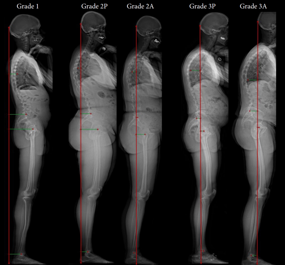
The PCVL was defined as a vertical plumb line from the posterior-most aspect of the occiput to the floor. The horizontal distances of the PCVL to the TA, posterior sagittal vertical line (PSVL; vertical line drawn superiorly from posterosuperior endplate of S1), the femoral head (FH) center, and center of the TP were measured. Each patient was then classified as either grade 1 (PCVL posterior to the TA and the PSVL), grade 2 (PCVL anterior to TA and posterior to the PSVL), or grade 3 (PCVL anterior to both the TA and the PSVL) (Fig. 1, Table 1). Grade 2 and 3 patients with a PCVL posterior to the center of the TP were given a “P” modifier; those with a PCVL anterior to the TP were given an “A” modifier (Table 1). All measurements were performed by a team of fellows and residents. A standardized protocol was created and written visual instructions were provided to every contributor. Individual measurements were reviewed throughout the cohort to assess interrater relatability using Cronbach alpha values, which exceeded 0.8 for all measures. Chi-square tests and analysis of variance were performed for bivariate analyses using SAS Studio Versions 3.4 (SAS Institute Inc., Cary, NC, USA).

RESULTS

Our study included a total of 334 asymptomatic volunteers, 64% of whom were female. Average age was 41±14 years and average BMI was 24.4 kg/m2. The average ODI score was 2.7±4.4 (Table 2). When measuring the relative position of the PCVL to various reference points, negative values were used to indicate a relatively posterior PCVL position, while a positive value indicated a relatively anterior PCVL position. Average PCVL–PSVL distance was -7.4 cm and average PCVL–TA distance was -2.1 cm. Average PCVL–FH distance was -10.8 cm and average PCVL–TP distance was -5.4 cm. Eighty-three percent of subjects were grade 1, 15% were grade 2, and 2% were grade 3. Overall, 98% of normal subjects were either grade 1 or grade 2.
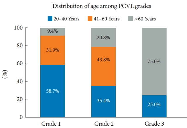
Higher PCVL grade had a significant association with increasing age (p<0.001). The mean age in grade 1 patients was 39.6 years, grade 2 was 45.6 years, and grade 3 was 63 years. Fig. 2 illustrates the age distribution of the asymptomatic cohort demonstrating an increase in age throughout the PCVL grades. Higher PCVL grade was also significantly associated with increasing greater T1 slope (p < 0.001), greater T4–12 kyphosis (p<0.001), greater cervical lordosis (p=0.006), greater pelvic tilt (p<0.001), and increased knee flexion (p<0.001) (Table 2). Conversely, higher PCVL grade was associated with decreased sacral slope (p=0.045). Higher PCVL grade was associated with a larger C7–S1 SVA (p<0.001) and C2–7 SVA (p<0.001) (Table 2). Of note, PI remained similar between PCVL grades as expected. A strong positive correlation (coefficient ≥ 0.6) was seen with C2–7 SVA and greater T1 slope, while a moderate correlation (coefficient ≥ 0.4) was seen with C7–S1 SVA and T4–12 thoracic kyphosis. Similarly, a PCVL anterior to the TP was strongly correlated with greater C7–S1 SVA (p < 0.001), C2–7 SVA (p<0.001), T1 slope (p<0.001) (Table 2).
When the population was subdivided using the TP modifier (anterior vs. posterior position of the PCVL relative to TP), similar results were seen with the anterior modifier and increasing PCVL grade. An anterior modifier had significant association with age (p=0.04) as well as increased C7–S1 SVA, increased C2–7 SVA, as well as increased T1 slope (p<0.001) (Table 3).

DISCUSSION

Dubousset first described “the cone of economy” decades ago—the upright posture of the human body that minimizes energy expenditure [23]. As sagittal alignment increases, energy expenditure increases as well to remain upright and maintain horizontal gaze. Compensation through the lumbar spine and pelvis occurs through pelvic retroversion—as lumbar lordosis decreases, PT increases and SS decreases [24]. In addition, there is an active, dynamic component maintaining this optimal position termed the chain of balance [15]. This complex anatomic relationship between the spine and pelvis has been well described by Roussouly et al. [25,26] as well, and the importance of sagittal spinal alignment in patient-reported outcomes has been well established previously [4,6-9].
The current widely accepted radiographic measurement for sagittal alignment, the C7–S1 SVA, has been considered the strongest predictor of improved outcomes in patients with ASD [4,22,27]. Lafage et al. [28] showed an SVA > 50 mm or PT > 20° were associated with worse outcomes scores. In a normal population, Gelb et al. [17] found the mean SVA to be -3.2±3.2 cm. The C7–S1 SVA, however, fails to the position of the skull and alignment of the cervical spine, as well as compensation through the lower extremities.
Various studies have shown that age-related changes and increasing positive sagittal alignment leads to compensatory knee flexion and subsequent hip extension [29-32]. Age-related changes have also been described in the cervical spine showing increasing C2–7 lordosis in order to maintain horizontal gaze [33,34]. These changes were also shown in a prior publication performed on a subset of the healthy volunteer radiographs used in this study [35]. Given these other compensatory mechanisms, it is not surprising that the focal C7–S1 SVA measurement is discordant with the gold standard GL (obtained using a force plate) [20-22].
The goal of our study was to develop a simple, practical radiographic measurement that could take into consideration all aspects of global sagittal alignment. A prior study by Kim et al. [36] attempted to address this issue with the cranial SVA, requiring a measurement from the midpoint of the nasion-inion line, from the root of the nose to the external occipital protuberance. While this did address skull position, we hoped to improve on this concept with a simple, practical radiographic marker that was easier to measure and reproduce reliably, with an organic anatomic grading system in the asymptomatic population. The PCVL requires a single vertical line drawn from the posterior-most aspect of the occiput down to the floor. In our study, the vast majority of healthy volunteers examined (98%) had a PCVL posterior to the PSVL (grades 1 and 2), with 83% of patients having a PCVL posterior to both the PSVL and TA (grade 1).
Aside from linear measurements such as the C7–S1 SVA, angular measurements have also been proposed to characterize global sagittal alignment. The cervicothoracic pelvic angle (CTPA), introduced by Protopsaltis et al. [37] in 2017, is defined as the angle formed between a line from C2 midvertebral body to the FH center and a line from T1 midvertebral body to the FH center. The CTPA has been shown to be associated with the development of proximal junctional kyphosis (PJK) postoperatively [37]. This is similar to the previously described T1PA, which is defined as the angle formed between a line from the T1 midvertebral body and the FH center to a line from the midpoint of the S1 superior endplate to the FH center [38]. T1PA has been shown to correlate with health-related quality of life for ASD patients [38].
In our study, we found that the PCVL grade does strongly correlate with the current gold standard C7–S1 SVA. In addition, we found that greater PCVL grade was significantly associated with expected compensatory changes over time such as increasing PI–LL, decreasing SS, increasing PT, increasing T1 slope. Regarding the skull and lower extremity position, the higher PCVL grade was significantly associated with expected compensatory increasing C2–7 lordosis, greater C2–7 SVA, as well as with increased knee flexion. In fact, C2–7 SVA and T1 slope both had a strong correlation (coefficient ≥ 0.6) with increasing PCVL grade. Based on our results, we hypothesize that the PCVL grade is an accurate reflection of global positive sagittal alignment, and as seen with prior sagittal measures that a higher PCVL grade may be associated with higher risk of PJK following a fusion procedure. The next step in this study would be to measure the PCVL in ASD patients preoperatively and postoperatively while tracking patient-reported outcome measures as well as radiographic evidence of PJK postoperatively.
When interpreting the results of this study, it is important to consider its limitations. First, the generalizability of the asymptomatic volunteers used in this study and whether they represent a true normal distribution of humanity upon which to base a new classification system may be brought into question. However, this cohort of asymptomatic volunteers is one of the largest collection of volunteers with full-standing radiographs across a wide variety of ages and ethnic backgrounds which we believe is a powerful tool in understanding a normative population. Secondly, the use of the PCVL requires radiographs to be obtained via imaging with 3-dimensional capabilities (EOS Imaging) which may not be available to every institution. Nonetheless, we believe that full-standing radiographs will be the new gold standard moving forward. Thus, this measure is crucial to further characterizing sagittal alignment with additional information provided by both the position of the skull as well as the lower extremities. Thirdly, the conducted study focuses on establishing a classification to patients with no spinal pathologies which may question if the proposed measurements correlate with the disease process. It is important to note that the purpose of this study is to establish a baseline distribution in the asymptomatic population in order to first characterize the measurement. Thus, the aforementioned relationship will be assessed in a subsequent study on its application in the ASD population.

CONCLUSION

We present the PCVL as a novel singular sagittal alignment marker that may play a helpful role in understanding global sagittal alignment. The PCVL is easy to implement and allows for immediate visualization of skull position relative to the thoracic spine, sacrum, as well as the TPs that is simple to interpret. Our population showed that a majority of asymptomatic volunteers were grade 1 (83%), while 17% of patients were grade 2 or 3 with a PCVL anterior to the TA. We propose here a simple grading system that may have the potential to aid in preoperative planning to correct sagittal malalignment, as well as assess patients postoperatively over time.

NOTES

Conflict of Interest

EOS imaging supported the multicentric collaborative group by providing the 3D parameters for the MEANS cohort and a web-based solution for image review. Authors, or their institutions did not receive any funding from EOS imaging relative to this work.

Funding/Support

This study received no specific grant from any funding agency in the public, commercial, or not-for-profit sectors.

Author Contribution

Conceptualization: PJP, ZMS, MPK, SB, KH, HKW, HWDH, HR, JCLH, LGL; Formal Analysis: PJP, FMH, MC; Investigation: LGL; Methodology: PJP, XEF, FMH, CM, NJL; Project Administration: FMH; Writing – Original Draft: PJP, XEF, FMH, CM, NJL, MC; Writing – Review & Editing: PJP, FMH, ZMS, MPK, SB, KH, HKW, HWDH, HR, JCLH, LGL.

Fig. 1.
The posterior cranial vertical line (PCVL) grading system. Grade 1: The PCVL lies posterior to the thoracic apex (TA) and posterior sagittal vertical line (PSVL). Grade 2: The PCVL lies posterior anterior to the TA and posterior to the PSVL. Grade 3: The PCVL lies anterior to both the TA and PSVL. The “A” and “P” modifiers refer to the PCVL position relative to the center of the tibial plafond in grades 2 and 3.
ns-2346408-204f1.jpg
Fig. 2.
Distribution of age among the posterior cranial vertical line (PCVL) grades. Grade 1: The PCVL lies posterior to the thoracic apex (TA) and posterior sagittal vertical line (PSVL). Grade 2: The PCVL lies posterior anterior to the TA and posterior to the PSVL. Grade 3: The PCVL lies anterior to both the TA and PSVL.
ns-2346408-204f2.jpg
Table 1.
The PCVL grading system with tibial plafond modifier
Grade Modifier PCVL position relative to
TA PSVL TP
Grade 1 (83%) - Posterior Posterior Posterior
Grade 2 (15%) P Anterior Posterior Posterior
A Anterior Posterior Anterior
Grade 3 (2%) P Anterior Anterior Posterior
A Anterior Anterior Anterior

PSVL, posterior sagittal vertical line; TA, thoracic apex; TP, tibial plafond.

Grade 1: The PCVL lies posterior to the thoracic apex (TA) and posterior sagittal vertical line (PSVL). Grade 2: The PCVL lies posterior anterior to the TA and posterior to the PSVL. Grade 3: The PCVL lies anterior to both the TA and PSVL. The “A” and “P” modifiers refer to the PCVL position relative to the center of the tibial plafond in grades 2 and 3.

Table 2.
The PCVL grade with demographic data and radiographic measurements
Variable PCVL grade
Correlation coefficient p-value
1 (N=276)
2 (N=48)
3 (N=8)
Mean ± SD Range Mean ± SD Range Mean ± SD Range
Age 39.6 ± 13.5 20.0–79.0 45.6 ± 14.9 20.0–76.0 63 ± 18.3 33.0–77.0 0.33 < 0.001*
BMI 24.2 ± 5.2 14.5–41.3 25.2 ± 5.4 16.9–44.7 26.4 ± 5.7 18.4–33.3 0.12 0.283
Knee flexion -2.4 ± 5 -22.3–17.7 -0.3 ± 4.6 -11.9–11.6 4.7 ± 5.7 -3.9–10.9 0.33 < 0.001*
C2–7 lordosis -1.3 ± 12.7 -31.4–43.3 3.3 ± 10.3 -14.9–27.8 8.9 ± 10.6 -13.6–17.1 0.26 0.006*
T4–12 kyphosis 34.9 ± 10.2 7.4–61.1 46.5 ± 11.7 27.5–77.8 49.8 ± 15 28.4–72.1 0.52 < 0.001*
Sacral slope 39.7 ± 8.3 13.6–65.6 37.5 ± 9.5 17.7–63.4 33.4 ± 11.6 17.0–52.1 -0.18 0.045*
Pelvic incidence 51.9 ± 10.5 27.3–81.8 50.1 ± 11.7 26.9–82.6 57 ± 11.5 38.8–68.2 -0.01 0.214
Pelvic tilt 12.3 ± 7.3 -6.9–41.9 12.6 ± 6.8 -3.5–27.3 23.6 ± 9.4 15.0–45.1 0.19 < 0.001*
PI–LL 9.4 ± 7.5 0.0–39.7 10.5 ± 7.7 0.2–28.1 10.5 ± 8.4 1.2–20.6 0.07 0.617
L1–S1 LL 56.8 ± 11.8 24.4–84.5 57.9 ± 12.8 25.3–86.1 47 ± 13.3 18.3–59.7 -0.06 0.060
C7–S1 SVA -9.3 ± 24.5 -77.1–56.9 5.7 ± 24.2 -41.0–54.1 67.9 ± 32.3 29.6–132.8 0.51 < 0.001*
C2–7 SVA 17.2 ± 8.6 -9.3–38.8 30.4 ± 8.9 12.9–53.2 30.4 ± 8.9 21.2–46.0 0.66 < 0.001*
T1 slope 21.4 ± 6.9 3.8–40.9 30.6 ± 6.4 20.8–52.1 38.8 ± 7.2 29.0–51.4 0.69 < 0.001*
ODI score 2.5 ± 4.3 0–20 3.5 ± 4.9 0–17.8 3.8 ± 4.9 0–14.0 0.12 0.261

PCVL, posterior cranial vertical line; SD, standard deviation; BMI, body mass index; PI–LL, pelvic incidence minus lumbar lordosis; SVA, sagittal vertical axis; ODI, Oswestry Disability Index.

Grade 1: The PCVL lies posterior to the thoracic apex (TA) and posterior sagittal vertical line (PSVL). Grade 2: The PCVL lies posterior anterior to the TA and posterior to the PSVL. Grade 3: The PCVL lies anterior to both the TA and PSVL.

Table 3.
Tibial plafond modifier with demographic data and radiographic measurements
Variable Posterior (N = 313) Anterior (N = 19) p-value
Age (yr) 40.7 ± 14.1 47.6 ± 18.2 0.040*
Body mass index (kg/m2) 24.5 ± 5.3 22.9 ± 3.9 0.226
Knee flexion -2.0 ± 5.1 -0.9 ± 5.2 0.353
C2–7 lordosis -0.5 ± 11 -0.4 ± 12.5 0.982
T4–12 kyphosis 36.6 ± 11.5 42.4 ± 9.6 0.034*
Sacral slope 39.1 ± 8.6 40.5 ± 9.9 0.516
Pelvic incidence 51.6 ± 10.6 53.9 ± 13.2 0.376
Pelvic tilt 12.5 ± 7.4 13.4 ± 8.0 0.599
PI–LL 9.6 ± 7.6 9.0 ± 6.8 0.741
L1–S1 LL 56.8 ± 12.1 56.1 ± 11.9 0.828
C7–S1 SVA -7.6 ± 25.4 33.7 ± 33.9 < 0.001*
C2–7 SVA 18.8 ± 9.7 29.9 ± 7.9 < 0.001*
T1 slope 22.6 ± 7.6 32.5 ± 7.9 < 0.001*
ODI score 2.6 ± 4.3 3.5 ± 5.2 0.375

Values are presented as mean±standard deviation.

PI–LL, pelvic incidence minus lumbar lordosis; SVA, sagittal vertical axis; ODI, Oswestry Disability Index.

* p<0.05, statistically significant differences.

REFERENCES

1. Eguchi Y, Suzuki M, Yamanaka H, et al. Influence of skeletal muscle mass and spinal alignment on surgical outcomes for lumbar spinal stenosis. Asian Spine J 2018;12:556-62.
crossref pmid pmc pdf
2. Gutman G, Labelle H, Barchi S, et al. Normal sagittal parameters of global spinal balance in children and adolescents: a prospective study of 646 asymptomatic subjects. Eur Spine J 2016;25:3650-7.
crossref pmid pdf
3. Iyer S, Sheha E, Fu MC, et al. Sagittal spinal alignment in adult spinal deformity: an overview of current concepts and a critical analysis review. JBJS Rev 2018;6:e2.
crossref
4. Glassman SD, Bridwell K, Dimar JR, et al. The impact of positive sagittal balance in adult spinal deformity. Spine (Phila Pa 1976) 2005;30:2024-9.
crossref pmid
5. Duval-Beaupere G, Schmidt C, Cosson P. A Barycentremetric study of the sagittal shape of spine and pelvis: the conditions required for an economic standing position. Ann Biomed Eng 1992;20:451-62.
crossref pmid pdf
6. Cho KJ, Suk SI, Park SR, et al. Risk factors of sagittal decompensation after long posterior instrumentation and fusion for degenerative lumbar scoliosis. Spine (Phila Pa 1976) 2010;35:1595-601.
crossref pmid
7. Glassman SD, Berven S, Bridwell K, et al. Correlation of radiographic parameters and clinical symptoms in adult scoliosis. Spine (Phila Pa 1976) 2005;30:682-8.
crossref pmid
8. Kim YJ, Bridwell KH, Lenke LG, et al. An analysis of sagittal spinal alignment following long adult lumbar instrumentation and fusion to L5 or S1: can we predict ideal lumbar lordosis? Spine (Phila Pa 1976) 2006;31:2343-52.
crossref pmid
9. Smith JS, Bess S, Shaffrey CI, et al. Dynamic changes of the pelvis and spine are key to predicting postoperative sagittal alignment after pedicle subtraction osteotomy: a critical analysis of preoperative planning techniques. Spine (Phila Pa 1976) 2012;37:845-53.
pmid
10. Mac-Thiong JM, Roussouly P, Berthonnaud E, et al. Age- and sex-related variations in sagittal sacropelvic morphology and balance in asymptomatic adults. Eur Spine J 2011;20 Suppl 5(Suppl 5):572-7.
crossref pmid pdf
11. Zhu Z, Xu L, Zhu F, et al. Sagittal alignment of spine and pelvis in asymptomatic adults: norms in Chinese populations. Spine (Phila Pa 1976) 2014;39:E1-6.
pmid
12. Le Huec JC, Saddiki R, Franke J, et al. Equilibrium of the human body and the gravity line: the basics. Eur Spine J 2011;20 Suppl 5(Suppl 5):558-63.
pmid
13. Gangnet N, Pomero V, Dumas R, et al. Variability of the spine and pelvis location with respect to the gravity line: a three-dimensional stereoradiographic study using a force platform. Surg Radiol Anat 2003;25:424-33.
crossref pmid pdf
14. Lafage V, Schwab F, Skalli W, et al. Standing balance and sagittal plane spinal deformity: analysis of spinopelvic and gravity line parameters. Spine (Phila Pa 1976) 2008;33:1572-8.
pmid
15. Hasegawa K, Okamoto M, Hatsushikano S, et al. Standing sagittal alignment of the whole axial skeleton with reference to the gravity line in humans. J Anat 2017;230:619-30.
crossref pmid pmc pdf
16. Van Royen BJ, Toussaint HM, Kingma I, et al. Accuracy of the sagittal vertical axis in a standing lateral radiograph as a measurement of balance in spinal deformities. Eur Spine J 1998;7:408-12.
crossref pmid pmc pdf
17. Gelb DE, Lenke LG, Bridwell KH, et al. An analysis of sagittal spinal alignment in 100 asymptomatic middle and older aged volunteers. Spine (Phila Pa 1976) 1995;20:1351-8.
crossref pmid
18. Hammerberg EM, Wood KB. Sagittal profile of the elderly. J Spinal Disord Tech 2003;16:44-50.
crossref pmid
19. McLean IP, Gillan MG, Ross JC, et al. A comparison of methods for measuring trunk list. A simple plumbline is the best. Spine (Phila Pa 1976) 1996;21:1667-70.
pmid
20. Yagi M, Takeda K, Machida M, et al. Discordance of gravity line and C7PL in patient with adult spinal deformity--factors affecting the occiput-trunk sagittal discordance. Spine J 2015;15:213-21.
crossref pmid
21. Roussouly P, Gollogly S, Noseda O, et al. The vertical projection of the sum of the ground reactive forces of a standing patient is not the same as the C7 plumb line: a radiographic study of the sagittal alignment of 153 asymptomatic volunteers. Spine (Phila Pa 1976) 2006;31:E320-5.
pmid
22. Mac-Thiong JM, Transfeldt EE, Mehbod AA, et al. Can c7 plumbline and gravity line predict health related quality of life in adult scoliosis? Spine (Phila Pa 1976) 2009;34:E519-27.
crossref pmid
23. Schwab F, Patel A, Ungar B, et al. Adult spinal deformity-postoperative standing imbalance: how much can you tolerate? An overview of key parameters in assessing alignment and planning corrective surgery. Spine (Phila Pa 1976) 2010;35:2224-31.
pmid
24. Savage JW, Patel AA. Fixed sagittal plane imbalance. Global Spine J 2014;4:287-96.
crossref pmid pmc pdf
25. Roussouly P, Pinheiro-Franco JL. Biomechanical analysis of the spino-pelvic organization and adaptation in pathology. Eur Spine J 2011;20 Suppl 5(Suppl 5):609-18.
crossref pmid pdf
26. Roussouly P, Gollogly S, Berthonnaud E, et al. Classification of the normal variation in the sagittal alignment of the human lumbar spine and pelvis in the standing position. Spine (Phila Pa 1976) 2005;30:346-53.
crossref pmid
27. Schwab FJ, Smith VA, Biserni M, et al. Adult scoliosis: a quantitative radiographic and clinical analysis. Spine (Phila Pa 1976) 2002;27:387-92.
pmid
28. Lafage V, Schwab F, Patel A, et al. Pelvic tilt and truncal inclination: two key radiographic parameters in the setting of adults with spinal deformity. Spine (Phila Pa 1976) 2009;34:E599-606.
pmid
29. Gottipati P, Fatone S, Koski T, et al. Crouch gait in persons with positive sagittal spine alignment resolves with surgery. Gait Posture 2014;39:372-77.
crossref pmid
30. Gottipati P, Stine R, Ganju A, et al. The effect of positive sagittal spine balance and reconstruction surgery on standing balance. Gait Posture 2018;62:227-34.
crossref pmid
31. Hasegawa K, Okamoto M, Hatsushikano S, et al. Normative values of spino-pelvic sagittal alignment, balance, age, and health-related quality of life in a cohort of healthy adult subjects. Eur Spine J 2016;25:3675-86.
crossref pmid pdf
32. Le Huec JC, Charosky S, Barrey C, et al. Sagittal imbalance cascade for simple degenerative spine and consequences: algorithm of decision for appropriate treatment. Eur Spine J 2011;20 Suppl 5(Suppl 5):699-703.
pmid
33. Park MS, Moon SH, Lee HM, et al. The effect of age on cervical sagittal alignment: normative data on 100 asymptomatic subjects. Spine (Phila Pa 1976) 2013;38:E458-63.
pmid
34. Yukawa Y, Kato F, Suda K, et al. Age-related changes in osseous anatomy, alignment, and range of motion of the cervical spine. Part I: Radiographic data from over 1,200 asymptomatic subjects. Eur Spine J 2012;21:1492-8.
crossref pmid pmc pdf
35. Iorio J, Lafage V, Lafage R, et al. The effect of aging on cervical parameters in a normative North American population. Global Spine J 2018;8:709-15.
crossref pmid pmc pdf
36. Kim YC, Lenke LG, Lee SJ, et al. The cranial sagittal vertical axis (CrSVA) is a better radiographic measure to predict clinical outcomes in adult spinal deformity surgery than the C7 SVA: a monocentric study. Eur Spine J 2017;26:2167-75.
crossref pmid pdf
37. Protopsaltis T, Bronsard N, Soroceanu A, et al. Cervical sagittal deformity develops after PJK in adult thoracolumbar deformity correction: radiographic analysis utilizing a novel global sagittal angular parameter, the CTPA. Eur Spine J 2017;26:1111-20.
crossref pmid pdf
38. Protopsaltis T, Schwab F, Bronsard N, et al. TheT1 pelvic angle, a novel radiographic measure of global sagittal deformity, accounts for both spinal inclination and pelvic tilt and correlates with health-related quality of life. J Bone Joint Surg Am 2014;96:1631-40.
crossref pmid


Editorial Office
CHA University, CHA School of Medicine Bundang Medical Center
59 Yatap-ro, Bundang-gu, Seongnam-si, Gyeonggi-do 13496, Korea
Tel: +82-31-780-1924  Fax: +82-31-780-5269  E-mail: support@e-neurospine.org
The Korean Spinal Neurosurgery Society
#407, Dong-A Villate 2nd Town, 350 Seocho-daero, Seocho-gu, Seoul 06631, Korea
Tel: +82-2-585-5455  Fax: +82-2-2-523-6812  E-mail: ksns1987@gmail.com
Business License No.: 209-82-62443

Copyright © The Korean Spinal Neurosurgery Society.

Developed in M2PI

Close layer
prev next