Warning: mkdir(): Permission denied in /home/virtual/lib/view_data.php on line 81 Warning: fopen(/home/virtual/e-kjs/journal/upload/ip_log/ip_log_2024-04.txt): failed to open stream: No such file or directory in /home/virtual/lib/view_data.php on line 83 Warning: fwrite() expects parameter 1 to be resource, boolean given in /home/virtual/lib/view_data.php on line 84 Comparative Efficacy of Surgical Interventions for Osteoporotic Vertebral Compression Fractures: A Systematic Review and Network Meta-analysis
Neurospine Search

CLOSE


Liu, Liu, Suvithayasiri, Han, and Kim: Comparative Efficacy of Surgical Interventions for Osteoporotic Vertebral Compression Fractures: A Systematic Review and Network Meta-analysis

Abstract

Objective

We aimed to comprehensively compare surgical methods for osteoporotic vertebral compression fracture (OVCF) using systematic review and network meta-analysis to understand their effectiveness and outcomes, as current research provides limited overviews.

Methods

We followed PRISMA (preferred reporting items for systematic reviews and meta-analyses) guidelines, preregistering our protocol with PROSPERO. We analyzed English-published randomized controlled trials (RCTs) on adults with OVCFs that evaluated pain intensity or functionality using tools like visual analogue scale (VAS) or Oswestry Disability Index (ODI). Exclusions included non-RCTs, malignancy-related fractures, and certain interventions. Using the RoB 2 tool, we assessed bias and visualized results with Robvis. Our primary outcome was pain intensity, with secondary outcomes including disability, new fractures, and cement leakage. Results were synthesized using Stata/MP.

Results

Thirty-four RCTs from 10 countries, totaling 4,384 patients, were analyzed. Short-term VAS indicated kyphoplasty with facet joint injection (KIJ) as the top treatment at 87.7%, while unipedicular kyphoplasty (UKP) led to long-term at 74.9%. Short-term ODI favored vertebroplasty with facet joint injection (VIJ) at 98.4%, with kyphoplasty (KP) leading long-term at 66.0%. All surgical techniques were superior to conservative treatment. Vertebral augmentation devices reported the fewest new fractures and curved vertebroplasty had the least cement leakage. SUCRA (surface under the cumulative ranking) analyses suggested UKP and VIJ as top choices for postoperative pain relief, with VIJ excelling in postoperative disability improvement.

Conclusion

Our analysis evaluates 12 OVCF interventions, underscoring KIJ for short-term pain relief and VIJ and UKP for long-term efficacy. Notably, VIJ stands out in disability outcomes, emphasizing the need for comprehensive OVCF management.

INTRODUCTION

A pivotal linkage exists between osteoporosis and osteoporotic vertebral compression fractures (OVCFs). This association has garnered considerable attention as an escalating health challenge and has become even more pronounced, especially among women populations that are progressively aging [1,2]. Given the global demographic tilt towards an older population, the prevalence of OVCF is anticipated to soar. The European Commission’s projections paint a worrisome scenario with a forecasted 57% surge in OVCF incidences in the European Union in the forthcoming 5 decades [3-5]. Parallel trends are evident in the United States, highlighting the widespread nature of this health predicament [6]. On a broader scale, approximately 1.4 million individuals globally are clinically diagnosed with OVCF each year, leading to a spectrum of outcomes from debilitating physical constraints to significant economic strains [7].
OVCF, characterized by intense pain and spinal deformities, substantially hinders the patient’s overall daily life activities. Disturbingly, while as many as 25% of women aged over 50 and 40% over 80 possess OVCF annually, an alarming two-thirds of such cases often remain undiagnosed or unattended [8]. The consequences of these fractures span from intense physical discomfort and loss of mobility to a palpable deterioration in overall quality of life [5]. While a majority of OVCF patients find success in conventional treatments such as bed rest, pain management, and rehabilitative exercises [9], there is a fragment of those for whom surgical interventions become imperative, especially when earlier treatments fail or the progression of deformities, or neurological deficits [9]. Although the current literature demonstrates many studies investigating varied available OVCF interventional options, there is still a lack in terms of a holistic, encompassing investigation of all available techniques. Moreover, those studies, often confined to the pairwise methods in their methodology, pose a challenge to obtaining a comprehensive overview. Therefore, we aim to provide an expansive investigation, juxtaposing all current surgical interventions for OVCF to evaluate their relative efficacies and clinical outcomes. Leveraging the usefulness of systematic review and the recently developed network meta-analysis (NMA) protocols, we intend to shed light on this potentially important underexplored area in this research.

MATERIALS AND METHODS

1. Search Strategy and Eligibility Criteria

This study was reported in accordance with the preferred reporting items for systematic reviews and meta-analyses (PRISMA) guidelines [10]. The protocol for our systematic review and meta-analysis was preregistered with PROSPERO (Registration No. CRD42023472135). We conducted comprehensive searches across PubMed (MEDLINE), Web of Science, Embase, and the Cochrane Library, covering from database inception to September 2, 2023. Adhering to the Cochrane Handbook for Systematic Reviews of Interventions. During the study selection process, 2 investigators independently assessed each study’s title, abstract, and full text, with disagreements arbitrated by a third reviewer. YT and SS extracted data into Microsoft Excel using a predefined standardized template. The extraction encompassed a comprehensive range of clinical and methodological trial characteristics, as outlined in the protocol. In instances of data extraction disagreements, a third reviewer (JSK) facilitated consensus. Key data points extracted from eligible studies included author, identifier, journal, country, publication year, duration, center, interventions, control group, follow-up duration, participant count by gender, age, outcome measurements, and utilized instruments.
To be eligible for this research, included studies must meet the following criteria: they should be fully published randomized controlled trials (RCTs) in English that involve adult participants (≥18 years) diagnosed with OVCF. These studies should comprise a control group receiving conservative treatments (Cts) and an intervention group exposed to treatments. The study outcomes should encompass pain intensity or functioning status measured through certificated clinical measurement tools like the visual analogue scale (VAS) or Oswestry Disability Index (ODI). If there are multiple publications from the same dataset, we will consider the one with the most comprehensive data and the longest follow-up period. Conversely, studies will be excluded if they are not RCTs, trials, reviews, case studies, conference abstracts, meeting proceedings, and editorials; deal with vertebral compression fractures stemming from malignancy or trauma, such as Kümmell Disease; employ stem cell therapy or minor modified technique, like novel design opening cannula; Intravenous Zoledronic acid infusion as variable intervention accompany with vertebroplasty (VP) or kyphoplasty (KP).

2. Risk of Bias Assessment and Outcomes Measurements

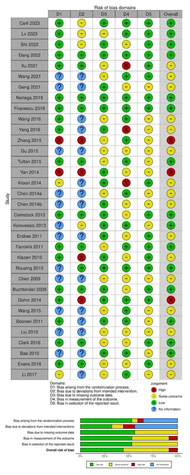
Two reviewers, YT and JL, independently evaluated the risk of bias (RoB) within the incorporated studies utilizing the RoB 2 tool specifically crafted for RCTs [11]. This robust assessment tool classifies biases into 5 distinct domains for individually randomized trials: (1) bias stemming from the randomization process, (2) bias resulting from deviations from the intended intervention, (3) bias owing to absent outcome data, (4) bias in the quantification of the outcome, and (5) bias in the selection of the reported result. For a more transparent and comprehensive depiction of the RoB evaluations, we employed the Robvis visualization tool to elucidate and present our findings in this systematic review and NMA [11].
The primary endpoint in our NMA model focused on pain intensity, gauged both short- and long-term through tools such as the VAS or Numerical Rating Scale. As for the secondary outcomes, they encompassed short- and long-term disability indices quantified through the ODI. Furthermore, the data took into account the number of new fractures and assessed instances of cement leakage during procedures involving cages.

3. Data Synthesis and Statistical Analysis

In our analysis, the NMA was executed using Stata software, taking advantage of the specialized “network” and “mvmeta” modules. We adopted a random-effects approach framed within the frequentist paradigm for aggregating the data extracted from RCTs. The software’s architecture presupposes that heterogeneity variance remains constant across all contrasted treatments within the study pool. A graphical network topology was generated to offer a visual depiction of the interrelationships among the selected studies [12]. In terms of research categorization, the studies were classified into Indirect Treatment Comparisons or Mixed Treatment Comparisons based on the emergent loop configurations within the network architecture. In cases of data unavailability, relevant statistical imputation techniques were employed. To ensure the reliability of the composite effect size, preliminary statistical assumptions were scrutinized to affirm the internal coherence of the NMA framework. To ascertain if heterogeneity originated from external or internal factors, node-splitting was conducted to evaluate the persistence of heterogeneity among the included studies. We computed effect sizes and their associated variances in direct and indirect pairwise comparisons, utilizing confidence and predictive intervals to adjust for varying levels of heterogeneity. We took additional steps to ascertain the uniformity of the aggregated data, applying logical inference hypotheses that account for potential inconsistencies.
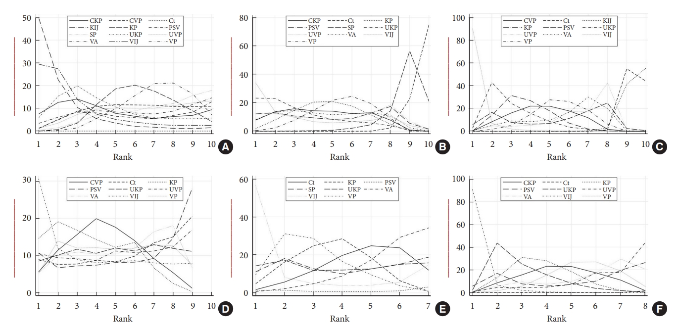
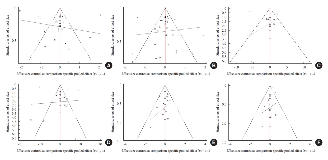
The net league square matrix is designed to encapsulate all pairwise comparisons in NMA. This methodology provides a comprehensive view of both direct and indirect evidence, enabling efficient interpretation of comparative treatment effects. Our analysis employed the surface under the cumulative ranking (SUCRA) methodology to formulate a treatment hierarchy. This method allowed for a data-driven, probabilistic stratification of the various therapeutic interventions and estimated the potential order of superiority for each clinical intervention by comparing their probability values against others [13]. SUCRA values range from 0% to 100%. Leveraging these SUCRA percentages, we executed a cluster analysis aimed at quantifying the holistic efficacy probabilities associated with each treatment option. Formal checks for publication bias were conducted through an asymmetrical analysis of comparison-adjusted funnel plots to spot any undue influence of smaller studies. All statistical computations and graphical renderings were facilitated using the comprehensive suite of network analysis tools available in Stata/MP (Release 17, StataCorp LLC, College Station, TX, USA).

RESULTS

1. Study Characteristics

The database search yielded 2,982 records, which were initially filtered using automation tools. A hands-on review of the titles and abstracts led to the exclusion of 138 articles that failed to satisfy the inclusion criteria. Consequently, we identified 34 RCTs spanning 10 countries, which collectively represented 4,384 patients, for inclusion in the NMA (Fig. 1). A detailed breakdown of the characteristics of these RCTs is provided in Table 1 [14-47]. Across these RCTs, 12 interventions were evaluated (Fig. 2), including VP, KP, unipedicular VP or KP (UVP or UKP), VP or KP with subsequent facet block or medial-branch-block injection (VIJ or KIJ), curved VP or KP (CVP or CKP) technique, pedicle screw fixation/fusion with or without vertebral augmentation (PSV), vertebral augmentation devices (VA), Sham procedure (SP), or Ct. The average participant age was 73.36 years. A significant proportion of the studies (70.59%) were undertaken in the United States, Australia, or China between 2009 and 2023. A thorough risk analysis revealed that 44.11% of the studies exhibited a low RoB, while 3 studies had notable risks tied to their randomization and intervention methodologies (Fig. 3).

2. NMA and Ranking Probabilities

Within a sample of 4,049 participants from 29 RCTs, pooled results of short-term VAS showed that Ct as a reference, CKP (mean 3.52, 95% confidence interval [CI] 1.23 to 5.81), CVP (mean 3.25, 95% CI 1.69 to 4.81), KIJ (mean 4.91, 95% CI 2.55 to 7.28), and other measurements illustrate the nonsignificant difference of improvement. Moreover, no significant difference was found among other pairwise comparisons. Furthermore, the long-term VAS outcomes displayed a similar trend when set Ct as a reference, with the exception that the groups treated with UKP (mean, 1.09; 95% CI, 0.08–2.09), VP (mean, 0.76; 95% CI, 0.07–1.45), and KP (mean, 0.86; 95% CI, 0.04–1.67) demonstrated significantly greater improvements compared to the SP group. The results were observed in the pairwise forest plot or netleague square matrix (Supplementary File). The cumulative probability for efficacy in short-term VAS indicates a marked preference for KIJ at 87.7%, followed by CKP and other treatments. In a broader sample involving 33 RCTs and 4,107 participants, when evaluating long-term VAS, UKP led the chart at 74.9%, with VIJ and other treatments following closely. Notably, nearly all surgical and SPs showcased a pronounced reduction in short-term VAS in contrast to Cts, with no discernible statistical difference in short-term pain intensity across surgical interventions. The net league diagram in Fig. 4 and Supplement Material provides a visual representation of comprehensive head-to-head comparisons for all measured metrics among various interventions.
For the ODI, evaluations showed distinct probabilities for both short- and long-term outcomes. Analyzing data from 17 RCTs with 2,087 participants for short-term outcomes, pooled results of all available measurements illustrate significant differences when conservative as reference. Additionally, the short-term improvements in ODI were significantly greater in the VIJ group compared to those observed in the KP (mean, 7.31; 95% CI, 0.80–13.83), UVP (mean, 11.71; 95% CI, 2.25–21.17), VA (mean, 10.51; 95% CI, 0.73–20.29), and VP (mean, 9.11; 95% CI, 3.37–14.85) groups. For the long-term ODI, pooled results only showed KP (mean, 10.46; 95% CI, 3.52–17.40) and VP (mean, 8.69; 95% CI, 3.16–14.21) demonstrated significantly greater improvements compared to the Ct group. The VIJ emerged as the leading intervention for OVCFs with a 98.4% probability, followed by UKP at 77.3%, KP at 69%, and CVP at 59.2%. In an evaluation focusing on long-term outcomes with 19 RCTs and 2,219 participants, KP topped the list with a 66.0% probability, trailed by VIJ at 63.1%, CVP at 56.7%, and VP at 49.2%. Notably, every surgical intervention displayed a significant advantage over Ct. Specifically, VIJ exhibited pronounced benefits in enhancing postoperative short-term ODI compared to most other treatments, such as KP, CVP, VP, VA, and UVP. Nevertheless, for long-term ODI outcomes, statistical comparisons between various clinical interventions did not yield significant disparities.
In assessing the occurrence of new fractures, data was drawn from 17 RCTs involving 2,087 participants. Interestingly, no marked differences were observed in the rates of new fractures among the VIJ, VP, KP, UKP, SP, Ct, VA, and PSV groups (Supplementary File). The SUCRA plot further highlighted that the VA intervention exhibited the lowest propensity for new fractures. Contrastingly, the incidence of cement leakage, analyzed from another set of 17 RCTs with 2,075 participants, showcased pronounced leakage variances in KP (relative risk [RR], 1.91; 95% CI, 1.02–3.60) and VP (RR, 1.73; 95% CI, 1.01–2.98) groups compared to the CVP group, as well as in the comparison between VP and UVP (RR, 8.69; 95% CI, 1.03–2.67) group. In this context, the SUCRA plot revealed that the CVP intervention had the least likelihood of cement leakage.
The SUCRA plot, as presented in Fig. 5, systematically ranks the surgical interventions based on both absolute and cumulative effects. Insights drawn from the SUCRA cluster analyses, illustrated in Fig. 6, suggest that UKP and VIJ potentially offer optimal relief from postoperative pain, both in the short and long run. Moreover, when focusing on postoperative disability outcomes over both short and long durations, VIJ may emerge as the most favorable intervention.

3. Consistency Test and Heterogeneity Analysis

In our network analysis, we observed controlled consistency between different treatments in terms of VAS, ODI, new fracture, and cement leakage (p ≥ 0.05). Only several arms showed significant inconsistency between different treatments for VAS, ODI, and adverse events, those considered the potential sources of populations or surgical techniques across the included studies. According to our statistical analysis, the design-by-treatment interaction inconsistency model is fit with no evidence for inconsistency in each outcome measurement using the restricted maximum likelihood. The global inconsistency values were p = 0.521 for short-term VAS, p = 0.983 for long-term ODI, p = 0.931 for short-term ODI, p = 0.373 for long-term ODI, p = 0.575 for new fracture, p = 0.931 for cement leakage, respectively.
Node-splitting analysis revealed low statistical inconsistency between direct and indirect evidence for the assessed treatment comparisons, suggesting our NMA results for these comparisons are robust. The comparison-adjusted symmetrical funnel plots demonstrated promising results, indicating a lower likelihood of publication bias or small-study effects. Notably, a minimal number of studies were located outside the funnel’s boundaries, further reinforcing the overall reliability and robustness of our findings (Fig. 7). Furthermore, due to the limited number of loops present, the potential for loop inconsistency was diminished. Therefore, we did not evaluate loop inconsistency that was made based on the specific structure of our network.

DISCUSSION

In the present NMA, we undertook an assessment of the clinical outcomes stemming from 12 interventions used for OVCFs. These outcomes were delineated by their short-term and long-term results. The VAS was employed to measure pain intensity, while the ODI gauged the extent of disability, serving as an indirect indicator of patients’ overall quality of life. When considering short-term results using the VAS, KIJ was pre-eminent. For long-term results, however, UKP took the lead but was also closely followed by VIJ. These findings underscore the pivotal role of subsequent injection procedures in pain management, both in immediate and enduring terms, as corroborated by contemporary research [48,49]. Prior studies have pinpointed the posterior segment of the vertebral body—particularly the facet joints —as a primary source of pain in certain situations. Age-related degeneration within spinal tissues, compounded by fractures, may intensify this discomfort. Notably, while VP targets pain arising from vertebral fractures, it demonstrates diminished efficacy in addressing pain attributed to residual injuries typically associated with posterior spinal trauma. For pain relief, targeting these nerves via facet joint injections has been proven efficacious [50]. A pioneering study by Kim et al. [51] evaluated the impact of combining percutaneous VP with facet block across 612 vertebral levels of OVCF. They deduced that this combined approach is advantageous owing to its minimal risks, expedited procedure, and heightened probability of facilitating pain alleviation and rapid return to mobilization. A recent RCT juxtaposed the efficiency and safety of VP combined with a subsequent facet joint block against VP alone. Although both methods proved beneficial, the combined approach exhibited superior back pain alleviation in the short term [17]. This convergence with ODI outcomes corroborates our findings, positioning VIJ at the forefront or near the top of our analysis. Therefore, this also underscores the amplified efficacy of postoperative injections in OVCF cases. Additional comprehensive RCTs, paired with an evaluation of cost-effectiveness relative to alternative interventions, are both essential and warranted in the future.
Concerning complications and safety, the most frequent severe issues postintervention in OVCF patients are new fractures and cement leakage. While no discernible differences exist in the rates of new fractures across our studied interventions, VA intervention exhibited the lowest propensity for new fractures according to the SUCRA analysis. Tutton et al. [30] have demonstrated the safety and effectiveness of the novel vertebral augmentation system (Kiva system) for the treatment of OVCF and found that it was noninferior to balloon KP to relieve pain and improved the overall function of the patients with superiority toward cement leakage and the new fracture rates. However, from our study, there is a notable discrepancy in cement leakage rates between the CVP group and the VP or KP groups. Two potential explanations arise for this distinction, both possibly related to the unique curved technique employed in the featured studies. Firstly, the amount of cement injected in the CVP group might be less than what is typically utilized in the standard KP or VP methods. Additionally, by strategically injecting the cement at the anterior zone of the vertebra with the aid of a flexible guide curving from one pedicle side, the procedure not only augments load support but also offers the advantage of administering the cement more anteriorly than traditional methods. As a result, the risk of cement leakage from the posterior wall of the vertebral body, which could gravely compress neurological structures, is likely to be diminished. Echoing our findings, Shi et al. [16] executed a 1:1 RCT, contrasting traditional KP with the innovative CVP technique on 90 OVCF patients. Both groups evidenced roughly comparable improvements in clinical outcomes. However, the CVP group fared significantly better in terms of lessening radiation exposure, operative duration, and the volume of cement required for injection. Intriguingly, despite the absence of statistical significance, there is an observable lessen trend toward a cement leakage rate in the CVP group. Similar optimistic results have been reported in other research [15,19,20]. Future studies encompassing a broader participant base are essential. Such investigations could potentially underscore the superior efficacy of CVP, particularly regarding its decreased propensity for cement leakage.
Intriguingly, our analysis found that SP significantly reduced the short-term VAS compared to Ct, a decrease that was on par with other interventions. This suggests the potential influence of the placebo effect, particularly during the initial stages of treatment. Therefore, caution is advised when interpreting results from such studies. This observation aligns with findings from several preceding studies. Numerous reputable RCTs have indicated no substantial difference in primary endpoints ranging from 6 months to 2 years between VP and SP, inferring that VP may not offer significant advantages over SP [22,37,47,52,53]. It is essential to clarify that referencing the “placebo effect” does not insinuate that the associated therapeutic outcomes are illusory. Comprehensive research, especially related to pain, has conclusively demonstrated that the placebo response is a genuine neurobiological event capable of delivering clinically significant advantages [54]. Some perspectives on this matter posit that the continuation of such procedures in clinical practice is hard to justify [55]. Even if a substantial component of improved clinical outcomes can be attributed to the placebo effect, there is skepticism about whether the placebo response from VP is substantial enough to warrant the associated risks. This is also considering numerous studies that have lauded the merits of VP [35,40,42,56]. However, in our NMA, although SP was shown to be efficacious, its SUCRA ranking did not eclipse that of other treatments. This observation, consistent with previously postulated theories, suggests that while a placebo effect might be present, it may not be dominant enough to overshadow the merits of existing treatments. This further underscores the value of our study’s NMA approach, allowing a comprehensive simultaneous comparison of multiple interventions, thereby providing a deeper understanding of the relative efficacy and standing of each treatment modality.
More from our findings, it is evident that every interventional technique outperformed Ct in every interested outcome. Based on prior research, Ct often falls short in alleviating pain and enhancing mobility, particularly in instances where there is a continued loss of vertebral body height or an exacerbation of kyphotic deformity. Additionally, if not meticulously diagnosed and managed, Ct can exacerbate disability, provoke intolerable side effects from narcotic analgesics, and induce bone demineralization [57]. Amidst the rapidly evolving therapeutic landscape for OVCFs and the broader domain of osteoporosis, a wealth of innovative evidence has surfaced, illuminating prospective and promising treatment directions. A seminal study by Shim et al. [58], for instance, investigated the combined use of Wharton’s jelly-derived mesenchymal stem cells and teriparatide in treating OVCFs. Their phase I/IIa research revealed that this combined modality is not only feasible and safe but also exceptionally effective in enhancing bone structure and expediting fracture healing. This groundbreaking work emphasizes the significance of amalgamating stem cell therapy with existing treatment modalities, offering a compounded benefit that might be transformative for OVCF management. As we navigate the surgical strategies for OVCFs, it becomes paramount to incorporate these cutting-edge developments, given their potential to elevate patient outcomes, diminish recuperation periods, and possibly curtail complications associated with conventional modalities. Merging stem cell therapy with relatively contemporary anabolic agents like teriparatide could usher in a revolutionary phase in OVCF treatment, warranting attention for future investigations and clinical applications.
We prioritized a stringent focus on RCTs, aiming to offset the known placebo effects common to invasive treatments and to address the complexities present in open-label studies adeptly in our study. A notable observation from our research was the pronounced heterogeneity across study findings. This could be linked to evolving surgical techniques, differences in practitioner expertise, and subjective variations in pain thresholds among participants. An interesting demographic pattern was the marked presence of females in OVCF cases. Methodologically, our study displayed several commendable attributes, including a thorough search protocol, the integration of a RoB evaluation to diminish inherent biases, and the strategic application of SUCRA analysis to assess the superior intervention comprehensively.
Despite these strengths, some limitations exist and need to be mentioned. First, a palpable lack of standardized criteria for discerning types and definitions of cement leakage—whether into the disc, perivertebral veins, pulmonary system, or spinal canal —was observed. Such an absence of consensus leads researchers to establish their own benchmarks, potentially obfuscating risk factor computations, especially when a meta-analysis is unduly reliant on a specific study. The lack of a further classification of the nature of fractures, whether adjacent or subsequent, or how much the extent of the pedicle screw fixation levels, whether it was short or long segments, or adjunctly use the augmentation or not, were not investigated in detail. All of these may also add another layer of heterogeneity, complicating comparisons across studies. Second, our research, while extensive, did not provide a detailed classification of osteoporosis severity. Undoubtedly, treatment recommendations for vertebral fractures vary depending on whether the patient has acute, chronic, severe, or generalized osteoporosis. The underreporting of detailed patient characteristics and their respective protocols of treatment related to OVCF in the included studies altered the choices of treatment and could make it vary between studies. Moreover, information on fracture types, severity, and duration, which are pivotal for tailoring treatment modalities and predicting outcomes, was inconsistently documented. This lack of granularity limits the depth of our analysis and the generalizability of our findings. Future research is thus encouraged to delve deeper into these nuances. We also recommend that forthcoming studies embrace a uniform reporting framework, including registering each RCT and providing an openly accessible detailed protocol. Enriching reports with visual data and related values would enhance transparency and comprehension. Third, the SUCRA rankings, although structured, are not infallible. These rankings might be grounded in evidence of inconsistent quality, thus leading to potentially shaky conclusions. SUCRA tends to emphasize singular outcomes, sidelining the intricate dynamics inherent in multi-dimensional medical treatments. It does not factor in critical considerations such as cost implications, practitioner familiarity with specific treatments, or nuanced differences in treatment impact. Additionally, the role of random chance on treatment outcomes is overlooked, potentially skewing perceptions of their comparative effectiveness. Thus, our conclusions, while informed, should be interpreted with a measure of restraint, not as incontrovertible evidence. To validate the efficacy of the diverse interventions, more extensive multi-arm RCTs are still essential.

CONCLUSION

In conclusion, the recent NMA meticulously assesses the effectiveness and safety of 12 interventions for OVCF. The data underscores KIJ’s prominence in short-term pain relief, while VIJ and UKP excel in long-term efficacy. VIJ consistently stands out in disability outcomes, emphasizing the complex nature of OVCF management. The success rates of KIJ and VIJ further accentuate the significance of injection procedures in pain management, suggesting the need for holistic treatment approaches that address both symptoms and root causes of OVCF pain. However, while our analysis is comprehensive, it is not exempt from the typical limitations, particularly concerning standardization in reporting cement leakages and fracture classifications. Future studies should entail a nuanced categorization of osteoporosis severity to ensure more precise treatment outcomes. With the ever-evolving medical field, integrating innovative treatments like stem cell therapy into OVCF procedures might be the way forward, affirming the continuous pursuit of enhanced patient outcomes.

Supplementary Material

NOTES

Conflict of Interest

Jin-Sung Kim is a consultant of RIWOSpine, GmbH, Germany; Elliquence, LLC, USA; and Stöckli Medical AG, Switzerland. The other authors have no conflicts of interest to declare.

Funding/Support

This study received no specific grant from any funding agency in the public, commercial, or not-for-profit sectors.

Author Contribution

Conceptualization: YL, SS, IH, JSK; Formal analysis: YL, JL; Methodology: YL, JL; Project administration: IH, JSK; Visualization: IH, JSK; Writing - original draft: YL, SS, JL; Writing - review & editing: YL, SS, IH, JSK.

Fig. 1.
Flowchart illustrating the database search strategy implemented in the study.
ns-2346996-498f1.jpg
Fig. 2.
Network geometry of the network meta-analysis. Network meta-analysis diagrams. (A) Short-term VAS. (B) Long-term VAS. (C) Short-term ODI. (D) Long-term ODI. (E) New fracture. (F) Cement leakage. VAS, visual analogue scale; ODI, Oswestry Disability Index; KP, kyphoplasty; CKP, curved kyphoplasty; Ct, conservative treatment; SP, Sham procedure; PSV, pedicle screw fixation/fusion with or without vertebral augmentation; VIJ, vertebroplasty with facet joint injection; VP, vertebroplasty; UVP, unilateral vertebroplasty; CVP, curved vertebroplasty; KIJ, kyphoplasty with facet joint injection; VA, vertebral augmentation devices; UKP, unipedicular kyphoplasty.
ns-2346996-498f2.jpg
Fig. 3.
Risk of bias assessment of the included randomized controlled trial studies according to the revised Cochrane risk of bias assessment tool.
ns-2346996-498f3.jpg
Fig. 4.
Netleague square matrix from the network meta-analysis detailing all pairwise treatment comparisons. KP, kyphoplasty; CKP, curved kyphoplasty; Ct, conservative treatment; SP, Sham procedure; PSV, pedicle screw fixation/fusion with or without vertebral augmentation; VIJ, vertebroplasty with facet joint injection; VP, vertebroplasty; UVP, unilateral vertebroplasty; CVP, curved vertebroplasty; KIJ, kyphoplasty with facet joint injection; VA, vertebral augmentation devices; UKP, unipedicular kyphoplasty; LF, long-term follow-up; SF, short-term follow-up; N/A, not applicable.
ns-2346996-498f4.jpg
Fig. 5.
SUCRA (surface under the cumulative ranking) plots that rank surgical interventions based on absolute and cumulative probability of effect. (A) Short-term VAS. (B) Long-term VAS. (C) Short-term ODI. (D) Long-term ODI. (E) New fracture. (F) Cement leakage. VAS, visual analogue scale; ODI, Oswestry Disability Index; KP, kyphoplasty; CKP, curved kyphoplasty; Ct, conservative treatment; SP, Sham procedure; PSV, pedicle screw fixation/fusion with or without vertebral augmentation; VIJ, vertebroplasty with facet joint injection; VP, vertebroplasty; UVP, unilateral vertebroplasty; CVP, curved vertebroplasty; KIJ, kyphoplasty with facet joint injection; VA, vertebral augmentation devices; UKP, unipedicular kyphoplasty.
ns-2346996-498f5.jpg
Fig. 6.
Hierarchical plots displaying the ranking of intervention probability on the impact of pain (A) intensity and disability index (B). UKP, unipedicular kyphoplasty; VIJ, vertebroplasty with facet joint injection; CVP, curved vertebroplasty; VA, vertebral augmentation devices; KP, kyphoplasty; VP, vertebroplasty; UVP, unilateral vertebroplasty; PSV, pedicle screw fixation/fusion with or without vertebral augmentation; Ct, conservative treatment; SP, Sham procedure.
ns-2346996-498f6.jpg
Fig. 7.
Comparison-adjusted funnel plots for the network of interventions. (A) Short-term VAS. (B) Long-term VAS. (C) Shortterm ODI. (D) Long-term ODI. (E) New fracture. (F) Cement leakage. VAS, visual analogue scale; ODI, Oswestry Disability Index.
ns-2346996-498f7.jpg
Table 1.
List of the included studies
No. Study Identifier Journal Country Year Time Center Interventions Control Follow-up N/Female Age (yr) Outcomes measurements
1 Carli et al. [14] NCT 01963039 Radiology Netherlands 2023 May 2013 to June 2019 Single-center Vertebroplasty Sham placebo 1 day, 1 wk, 1, 3, 6, 12 mo 80/54 69 ± 10 VAS, QUALEFFO, RMDQ, analgesics use, fracture cleft, new fractures
2 Lv et al. [15] ChiCTR 2000036792 Spine China 2023 September 2020 to April 2022 Single-center Curved vertebroplasty Unipedicular vertebroplasty 1 day, 1 wk, 1, 3, 6, 12 mo 103/59 74.81 ± 5.87 VAS, ODI, cement leakage, cement distribution, cement volume, fluoroscopy times, operation duration
3 Shi et al. [16] ChiCTR 2200056785 The spine journal China 2022 May 2019 to October 2020 Single-center Curved kyphoplasty Bilateral kyphoplasty 1 day, 6, 12 mo 85/71 70.99 ± 6.79 VAS, ODI, anterior edge height, kyphosis angulation, cement distribution, cement leakage, leakage pattern, frequency of intraoperative fluoroscopy, operative time, cement volume
4 Dang et al. [17] ChiCTR 2200056526 BMC Musculoskeletal Disorders China 2022 January 2018 to December 2020 Single-center Vertebroplasty with facet joint block Vertebroplasty 1 day, 1, 3, 6, 12 mo 198/103 77.39 ± 7.78 VAS, ODI, cement volume, hospitalization time, operation time, cement volume complication rate, Cox-2 inhibitors
5 Xu et al. [18] SAHoWMU-CR2018-08-109 Pain Physician China 2021 August 2019 to November 2019 Single-center Kyphoplasty with medial branch block Kyphoplasty 1 day, 1 wk, 1, 3, 6, 12 mo 179/151 N/A VAS, PROMIS PF, satisfaction, the incidence of residual back pain
6 Wang et al. [19] ChiCTR 2100042859 BMC Musculoskeletal Disorders China 2021 January 2019 to January 2020 Single-center Curved vertebroplasty Kyphoplasty 1 day, 6 mo 72/50 76.04 ± 6.15 VAS, ODI, fluoroscopy times, cement volume, anterior edge height, Cobb angle, cement leakage
7 Geng et al. [20] N/A Orthopedics China 2021 February 2016 to February 2018 Single-center Curved vertebroplasty Vertebroplasty 2 day, 3 mo 96/64 70.56 ± 5.91 VAS, ODI, Cobb angle, vertebral body height variation
8 Noriega et al. [21] NCT02461810 The spine journal Spain 2019 April 2015 to February 2017 Multi-center Titanium vertebral augmentation kyphoplasty 5 day, 1, 6, 12 mo 141/111 73.3 ± 9.5 Pain intensity, BMD, ODI, EQ-5D, ambulatory status, analgesic consumption, length of hospital stay, symptomatic cement extravasation requiring surgical reintervention or retreatment
9 Firanescu et al. [22] NCT01200277 the BMJ Netherlands 2018 January 2011 to January 2013 Multi-center Vertebroplasty Sham placebo 1 day, 1 wk, 1, 3, 6, 12 mo 176/133 75.78 ± 9.56 VAS, QUALEFFO, RMDQ
10 Li et al. [23] N/A Int J Clin Exp Med China 2017 January 2013 to June 2015 Single-center Kyphoplasty Conservative 1 wk, 1, 3, 6 mo 80/24 74.03 ± 6.21 VAS, ODI, Cobb angle, vertebral height, vertebral fracture restoration, symptom relief
11 Wang et al. [24] N/A Eur Spine J China 2016 January 2009 and January 2013 Single-center Facet joint block Vertebroplasty 1 day, 1 wk, 1, 3, 6, 12 mo 200/165 63.14 ± 5.56 VAS, ODI, BMD, SF-36, new fractures
12 Yang et al. [25] N/A Spine China 2016 January 2009 to December 2011 Single-center Vertebroplasty Conservative treatment 1 day, 1 wk, 1, 3, 6, 12 mo 107/69 76.67 ± 5.80 VAS, ODI, QUALEFFO, satisfaction surveys
13 Clark et al. [26] NCT01482793 Lancet Australia 2016 Nov 2011 to Dec 2014 Multi-center Vertebroplasty Sham placebo 3, 14 day, 1, 3, 6 mo 120/88 80.49 ± 6.99 NRS, RMDQ, QUALEFFO, EQ-5D, analgesic use
14 Evans et al. [27] NCT00279877 Journal of NeuroInterventional Surgery United States 2016 N/A Multi-center Vertebroplasty Kyphoplasty 3, 30 day, 6, 12 mo 115/82 75.6 ± 10.0 VAS, RMDQ, SOF-6 ADL, EQ-5D, SF-36, OPAQ
15 Zhang et al. [28] N/A BMC Musculoskeletal Disorders China 2015 November 2010 to October 2012 Single-center Uni-vertebroplasty Bi-vertebroplasty 3 day, 24 mo 50/ 37 71.91 ± 6.69 VAS, ODI, SF-36, anterior/middle vertebral body height variation, Cement leakage, Cement volume, operation time, radiation time, incidence of complications
16 Gu et al. [29] N/A Journal of Orthopaedic Surgery and Research China 2015 November 2010 to August 2011 Single-center Pedicle screw fixation Vertebroplasty 1, 2, 3, 6, 12, 24 mo 68/47 74.55 ± 5.91 VAS, anterior/central vertebral body height, local kyphosis, Cobb angle
17 Tutton et al. [30] N/A Spine United States 2015 August 2010 to May 2013 Multi-center Kiva Augmentation Treatment Kyphoplasty 30 day, 6, 12 mo 285/211 75.56 ± 9.22 VAS, ODI, DXA, cement volume, cement extravasation, absence of device-related SAEs, adjacent fracture
18 Wang et al. [31] N/A Pain Physician China 2015 January 2012 to February 2014 Single-center Vertebroplasty Kyphoplasty 1 day, 3, 12 mo 107/81 69.03 ± 8.64 VAS, ODI, location of leakages, cement leakage, and vertebral height restoration, cement volumes
19 Yan et al. [32] N/A Spine China 2014 January 2009 to January 2012 Single-center Uni-kyphoplasty Bi-kyphoplasty 1, 3, 6, 12 mo 309/220 71.51 ± 3.98 VAS, SF-36, RMDQ, cement volume, radiation dose, anterior/posterior vertebral body height, kyphotic angle, cement distribution
20 Kroon et al. [33] ACTRN01260 5000079640 Journal of Bone and Mineral Research Australia 2014 April 2004 to October 2008 Multi-center Vertebroplasty Sham 1 wk, 1, 3, 6, 12, 24 mo 57/47 77.19 ± 9.23 Pain intensity, QUALEFFP, AQoL, RMDQ, EQ-5D, timed up & go, cement volume, pain at rest and at night, disability, quality of life, perceived recovery, adverse events
21 Chen et al. [34] N/A Journal of spinal disorders & techniques China 2014 N/A Single-center Uni-vertebroplasty Bi-vertebroplasty 2 wk, 24 mo 39/N/A 69.06 ± 7.49 VAS, ODI, cement volume, cement leakage
22 Chen et al. [35] N/A Journal of Clinical Neuroscience China 2014 January 2007 to December 2012 Single-center Vertebroplasty Conservative 1 wk, 1, 3, 6, 12 mo 89/62 65.53 ± 9.10 VAS, ODI, RMDQ, drugs control, pain relief, functional outcome
23 Dohm et al. [36] NCT00323609 Am J Neuroradiol United States 2014 October 2006 to May 2011 Multi-center Vertebroplasty Kyphoplasty 1 day, 3, 12, 24 mo 361/280 75.6 VAS, ODI, SF-36, EQ-5D, cement volume, cement leakage, radiographic fracture, new radiographic VCF, kyphotic angulation
24 Comstock et al. [37] NCT00068822 Neuroradiology United States 2013 June 2004 to August 2008 Multi-center Vertebroplasty Sham placebo 1, 3, 6, 12 mo 131/N/A N/A Pain intensity, RMDQ
25 Korovessis et al. [38] N/A J Bone & Mineral Research (Spine) Greece 2013 May 2010 to September 2010 Single-center Kiva Aug-mentation Treatment Kyphoplasty 12 mo 168/119 70.98 ± 10.34 VAS, SF-36, ODI, anterior/midline/posterior vertebra body height ratio, cement leakage, Gardner angle
26 Endres et al. [39] AZ 2010-218-f-s Orthopaedics & Traumatology: Surgery & Research Germany 2011 N/A Single-center Soteira shield kyphoplasty system Kyphoplasty/Vertebroplasty 6 mo 59/40 53–77 VAS, ODI, dose-area product, beck index, fluoroscopy times, radiological imaging with regard to height restoration, cement distribution, cement leakage, recurrent fracture
27 Farrokhi et al. [40] IRCT13880 4252193N1 J Neurosurg Spine Iran 2011 September 2004 to January 2006 Single-center Vertebroplasty Conservative 1 wk, 2, 6, 12, 24, 36 mo 82/60 55–90 VAS, ODI, vertebra body height, kyphotic angulation, sagittal index
28 Boonen et al. [41] N/A Journal of Bone and Mineral Research United Kingdom 2011 February 2003 to December 2005 Multi-center Kyphoplasty Conservative 7 day, 1, 3, 6, 12, 24 mo 300/232 44.5–95.2 VAS, SF-36, RMDQ, reduced activity, EQ-5D, limited days of activity and bed rest, patient satisfaction (20-point Likert scale)
29 Klazen et al. [42] NCT00232466 Lancet Netherlands 2010 October 2005 to June 2008 Multi-center Vertebroplasty Conservative 1 day, 1 wk, 1, 3, 6, 12 mo 202/140 75.30 ± 9.10 VAS, QUALEFFO, EQ-5D, RMDQ, medical costs, pain relief, cost-effectiveness, QALYs, RMDQ
30 Rousing et al. [43] 20000161-20030072 Spine Denmark 2010 January 2001 to January 2008 Single-center Vertebroplasty Conservative 3, 12 mo 49/40 65–96 VAS, SF-36, DPQ, EQ-5D, Barthel, MMSE, tandem test, timed up & go, chair test
31 Liu et al. [44] N/A Osteoporos Int China 2010 N/A Single-center Vertebroplasty Kyphoplasty 3 day, 6 mo 100/77 57–84 VAS, vertebral body height, kyphotic wedge angle, location of osteoporotic VCFs, period between injury and surgery
32 Bae et al. [45] N/A Spine United Kingdom 2010 N/A Multi-center Vertebroplasty Kyphoplasty 1 wk, 1, 3, 6, 12, 24, 36 mo 40/26 75.5 ± 12.15 VAS, ODI, SF-12, cement volume
33 Chen et al. [46] N/A Injury China 2009 N/A Single-center Uni-kyphoplasty Bi-kyphoplasty 2 wk 58/N/A 68.07 ± 7.09 VAS, ODI, restoration rate, surgery time, vertebral height changes, clinical outcomes
34 Buchbinder et al. [47] ACTRN0126050 00079640 N Engl J Med Australia 2009 April 2004 to October 2008 Multi-center Vertebroplasty Sham placebo 1 wk, 1, 3, 6 mo 78/62 76.61 ± 12.06 Pain intensity, QUALEFFP, AQoL, RMDQ, EQ-5D

VAS, visual analogue scale; QUALEFFO, quality of life questionnaire; RMDQ, Roland Morris Disability Questionnaire; ODI, Oswestry Disability Index; NRS, Numerical Rating Scale; PROMIS PF, patient-reported outcomes measurement information system physical function; BMD, bone mineral density; EQ-5D, European quality of life–5 dimensions; SF-36, 36-item Short Form health survey; SOF ADL, Study of Osteoporotic Fractures Index Scoring activities of daily living; OPAQ, Osteoporosis Assessment Questionnaire; DXA, dual-energy x-ray absorptiometry; AQoL, assessment of quality of life; VCF, vertebral compression fracture; QALYs, quality-adjusted life years; SF-12, 12-item Short Form health survey; DPQ, Dallas Pain Questionnaire; MMSE, modified mini-mental state examination.

REFERENCES

1. Peck WA. Consensus development conference: diagnosis, prophylaxis, and treatment of osteoporosis. Am J Med 1993;94:646-50.
crossref pmid
2. Kanis JA. Assessment of fracture risk and its application to screening for postmenopausal osteoporosis: synopsis of a WHO report. WHO Study Group. Osteoporos Int 1994;4:368-81.
pmid
3. Strom O, Borgstrom F, Zethraeus N, et al. Long-term cost and effect on quality of life of osteoporosis-related fractures in Sweden. Acta Orthop 2008;79:269-80.
crossref pmid
4. Borgstrom F, Zethraeus N, Johnell O, et al. Costs and quality of life associated with osteoporosis-related fractures in Sweden. Osteoporos Int 2006;17:637-50.
crossref pmid pdf
5. Kanis JA, Johnell O, Oden A, et al. The risk and burden of vertebral fractures in Sweden. Osteoporos Int 2004;15:20-6.
crossref pmid pdf
6. Cummings SR, Melton LJ. Epidemiology and outcomes of osteoporotic fractures. Lancet 2002;359:1761-7.
crossref pmid
7. Johnell O, Kanis JA. An estimate of the worldwide prevalence and disability associated with osteoporotic fractures. Osteoporos Int 2006;17:1726-33.
crossref pmid pdf
8. Silverman SL. The clinical consequences of vertebral compression fracture. Bone 1992;13 Suppl 2:S27-31.
crossref pmid
9. Kim DH, Vaccaro AR. Osteoporotic compression fractures of the spine; current options and considerations for treatment. Spine J 2006;6:479-87.
crossref pmid
10. Hutton B, Salanti G, Caldwell DM, et al. The PRISMA extension statement for reporting of systematic reviews incorporating network meta-analyses of health care interventions: checklist and explanations. Ann Intern Med 2015;162:777-84.
crossref pmid
11. Sterne JAC, Savović J, Page MJ, et al. RoB 2: a revised tool for assessing risk of bias in randomised trials. BMJ 2019;366:l4898.
crossref pmid
12. Salanti G, Kavvoura FK, Ioannidis JP. Exploring the geometry of treatment networks. Ann Intern Med 2008;148:544-53.
crossref pmid
13. Mbuagbaw L, Rochwerg B, Jaeschke R, et al. Approaches to interpreting and choosing the best treatments in network meta-analyses. Sys Rev 2017;6:79.
crossref pmid pmc pdf
14. Carli D, Venmans A, Lodder P, et al. Vertebroplasty versus active control intervention for chronic osteoporotic vertebral compression fractures: the VERTOS V randomized controlled trial. Radiology 2023;308:e222535.
crossref pmid
15. Lv Z, Chen Z, Chen H, et al. Percutaneous curved vertebroplasty versus unipedicular approach vertebroplasty for acute osteoporotic vertebral compression fractures : a randomized controlled trial. Spine (Phila Pa 1976) 2023;48:552-8.
pmid
16. Shi X, Li P, Li J, et al. Comparative evaluation of an innovative deflectable percutaneous kyphoplasty versus conventional bilateral percutaneous kyphoplasty for osteoporotic vertebral compression fractures: a prospective, randomized and controlled trial. Spine J 2023;23:585-98.
crossref pmid
17. Dang SJ, Wei WB, Wei L, et al. Vertebroplasty combined with facet joint block vs. vertebroplasty alone in relieving acute pain of osteoporotic vertebral compression fracture: a randomized controlled clinical trial. BMC Musculoskelet Disord 2022;23:807.
crossref pmid pmc pdf
18. Xu JJ, Tang XT, Yang J, et al. The effect of medial branch block on postoperative residual pain relieve after percutaneous kyphoplasty: a randomized controlled trial with 12-month follow-up. Pain Physician 2021;24:E1059-66.
pmid
19. Wang C, Zhang Y, Chen W, et al. Comparison of percutaneous curved kyphoplasty and bilateral percutaneous kyphoplasty in osteoporotic vertebral compression fractures: a randomized controlled trial. BMC Musculoskelet Disord 2021;22:588.
crossref pmid pmc pdf
20. Geng Z, Zhou Q, Shang G, et al. Short-term efficacy of the percutaneous vertebroplasty using a curved versus straight vertebroplasty needle in osteoporotic vertebral compression fractures. Orthopedics 2021;44:e131-8.
crossref pmid
21. Noriega D, Marcia S, Theumann N, et al. A prospective, international, randomized, noninferiority study comparing an implantable titanium vertebral augmentation device versus balloon kyphoplasty in the reduction of vertebral compression fractures (SAKOS study). Spine J 2019;19:1782-95.
crossref pmid
22. Firanescu CE, de Vries J, Lodder P, et al. Vertebroplasty versus sham procedure for painful acute osteoporotic vertebral compression fractures (VERTOS IV): randomised sham controlled clinical trial. BMJ 2018;361:k1551.
crossref pmid pmc
23. Li Y, Zhu J, Xie C. A comparative study of percutaneous kyphoplasty and conservative therapy on vertebral osteoporotic compression fractures in elderly patients. Int J Clin Exp Med 2017;10:8139-45.

24. Wang B, Guo H, Yuan L, et al. A prospective randomized controlled study comparing the pain relief in patients with osteoporotic vertebral compression fractures with the use of vertebroplasty or facet blocking. Eur Spine J 2016;25:3486-94.
crossref pmid pdf
25. Yang EZ, Xu JG, Huang GZ, et al. Percutaneous vertebroplasty versus conservative treatment in aged patients with acute osteoporotic vertebral compression fractures: a prospective randomized controlled clinical study. Spine (Phila Pa 1976) 2016;41:653-60.
pmid
26. Clark W, Bird P, Gonski P, et al. Safety and efficacy of vertebroplasty for acute painful osteoporotic fractures (VAPOUR): a multicentre, randomised, double-blind, placebo-controlled trial. Lancet 2016;388:1408-16.
crossref pmid
27. Evans AJ, Kip KE, Brinjikji W, et al. Randomized controlled trial of vertebroplasty versus kyphoplasty in the treatment of vertebral compression fractures. J Neurointerv Surg 2016;8:756-63.
crossref pmid
28. Zhang L, Liu Z, Wang J, et al. Unipedicular versus bipedicular percutaneous vertebroplasty for osteoporotic vertebral compression fractures: a prospective randomized study. BMC Musculoskelet Disord 2015;16:145.
crossref pmid pmc pdf
29. Gu YT, Zhu DH, Liu HF, et al. Minimally invasive pedicle screw fixation combined with percutaneous vertebroplasty for preventing secondary fracture after vertebroplasty. J Orthop Surg Res 2015;10:31.
crossref pmid pmc pdf
30. Tutton SM, Pflugmacher R, Davidian M, et al. KAST study: the kiva system as a vertebral augmentation treatment-a safety and effectiveness trial: a randomized, noninferiority trial comparing the Kiva System with balloon kyphoplasty in treatment of osteoporotic vertebral compression fractures. Spine (Phila Pa 1976) 2015;40:865-75.
pmid
31. Wang CH, Ma JZ, Zhang CC, et al. Comparison of high-viscosity cement vertebroplasty and balloon kyphoplasty for the treatment of osteoporotic vertebral compression fractures. Pain Physician 2015;18:E187-94.
pmid
32. Yan L, Jiang R, He B, et al. A comparison between unilateral transverse process-pedicle and bilateral puncture techniques in percutaneous kyphoplasty. Spine (Phila Pa 1976) 2014;39(26 Spec No.):B19-26.
crossref pmid
33. Kroon F, Staples M, Ebeling PR, et al. Two-year results of a randomized placebo-controlled trial of vertebroplasty for acute osteoporotic vertebral fractures. J Bone Miner Res 2014;29:1346-55.
crossref pmid
34. Chen C, Bian J, Zhang W, et al. Unilateral versus bilateral vertebroplasty for severe osteoporotic vertebral compression fractures. J Spinal Disord Tech 2014;27:E301-4.
crossref pmid
35. Chen D, An ZQ, Song S, et al. Percutaneous vertebroplasty compared with conservative treatment in patients with chronic painful osteoporotic spinal fractures. J Clin Neurosci 2014;21:473-7.
crossref pmid
36. Dohm M, Black CM, Dacre A, et al. A randomized trial comparing balloon kyphoplasty and vertebroplasty for vertebral compression fractures due to osteoporosis. AJNR Am J Neuroradiol 2014;35:2227-36.
crossref pmid pmc
37. Comstock BA, Sitlani CM, Jarvik JG, et al. Investigational vertebroplasty safety and efficacy trial (INVEST): patient-reported outcomes through 1 year. Radiology 2013;269:224-31.
crossref pmid pmc
38. Korovessis P, Vardakastanis K, Repantis T, et al. Balloon kyphoplasty versus KIVA vertebral augmentation--comparison of 2 techniques for osteoporotic vertebral body fractures: a prospective randomized study. Spine (Phila Pa 1976) 2013;38:292-9.
pmid
39. Endres S, Badura A. Shield kyphoplasty through a unipedicular approach compared to vertebroplasty and balloon kyphoplasty in osteoporotic thoracolumbar fracture: a prospective randomized study. Orthop Traumatol Surg Res 2012;98:334-40.
crossref pmid
40. Farrokhi MR, Alibai E, Maghami Z. Randomized controlled trial of percutaneous vertebroplasty versus optimal medical management for the relief of pain and disability in acute osteoporotic vertebral compression fractures. J Neurosurg Spine 2011;14:561-9.
crossref pmid
41. Boonen S, Van Meirhaeghe J, Bastian L, et al. Balloon kyphoplasty for the treatment of acute vertebral compression fractures: 2-year results from a randomized trial. J Bone Miner Res 2011;26:1627-37.
crossref pmid
42. Klazen CA, Lohle PN, de Vries J, et al. Vertebroplasty versus conservative treatment in acute osteoporotic vertebral compression fractures (Vertos II): an open-label randomised trial. Lancet 2010;376:1085-92.
crossref pmid
43. Rousing R, Hansen KL, Andersen MO, et al. Twelve-months follow-up in forty-nine patients with acute/semiacute osteoporotic vertebral fractures treated conservatively or with percutaneous vertebroplasty: a clinical randomized study. Spine (Phila Pa 1976) 2010;35:478-82.
pmid
44. Liu JT, Liao WJ, Tan WC, et al. Balloon kyphoplasty versus vertebroplasty for treatment of osteoporotic vertebral compression fracture: a prospective, comparative, and randomized clinical study. Osteoporos Int 2010;21:359-64.
crossref pmid pdf
45. Bae H, Shen M, Maurer P, et al. Clinical experience using Cortoss for treating vertebral compression fractures with vertebroplasty and kyphoplasty: twenty four-month follow-up. Spine (Phila Pa 1976) 2010;35:E1030-6.
pmid
46. Chen C, Chen L, Gu Y, et al. Kyphoplasty for chronic painful osteoporotic vertebral compression fractures via unipedicular versus bipedicular approachment: a comparative study in early stage. Injury 2010;41:356-9.
crossref pmid
47. Buchbinder R, Osborne RH, Ebeling PR, et al. A randomized trial of vertebroplasty for painful osteoporotic vertebral fractures. N Engl J Med 2009;361:557-68.
crossref pmid
48. Georgy BA. Interventional techniques in managing persistent pain after vertebral augmentation procedures: a retrospective evaluation. Pain Physician 2007;10:673-6.
crossref pmid
49. Lehman VT, Wood CP, Hunt CH, et al. Facet joint signal change on MRI at levels of acute/subacute lumbar compression fractures. AJNR Am J Neuroradiol 2013;34:1468-73.
crossref pmid pmc
50. Balazsfi M, Kis D, Toth T, et al. Radiofrequency facet joint denervation efficiency based on the severity of spondylarthrosis and in osteoporotic vertebral compression fractures. A retrospective study. Clin Neurol Neurosurg 2019;186:105497.
crossref pmid
51. Kim TK, Kim KH, Kim CH, et al. Percutaneous vertebroplasty and facet joint block. J Korean Med Sci 2005;20:1023-8.
crossref pmid pmc
52. Kallmes DF, Comstock BA, Heagerty PJ, et al. A randomized trial of vertebroplasty for osteoporotic spinal fractures. N Engl J Med 2009;361:569-79.
crossref pmid pmc
53. Staples MP, Howe BM, Ringler MD, et al. New vertebral fractures after vertebroplasty: 2-year results from a randomised controlled trial. Arch Osteoporos 2015;10:229.
crossref pmid pmc pdf
54. Benedetti F. Placebo effects: understanding the mechanisms in health and disease. Oxford (England): Oxford University Press; 2009.

55. Miller FG, Kallmes DF, Buchbinder R. Vertebroplasty and the placebo response. Radiology 2011;259:621-5.
crossref pmid pmc
56. Blasco J, Martinez-Ferrer A, Macho J, et al. Effect of vertebroplasty on pain relief, quality of life, and the incidence of new vertebral fractures: a 12-month randomized follow-up, controlled trial. J Bone Miner Res 2012;27:1159-66.
crossref pmid
57. Klazen CA, Verhaar HJ, Lohle PN, et al. Clinical course of pain in acute osteoporotic vertebral compression fractures. J Vasc Interv Radiol 2010;21:1405-9.
crossref pmid
58. Shim J, Kim KT, Kim KG, et al. Safety and efficacy of Wharton’s jelly-derived mesenchymal stem cells with teriparatide for osteoporotic vertebral fractures: a phase I/IIa study. Stem Cells Transl Med 2021;10:554-67.
crossref pmid pmc pdf
  • Medtronic
  • TOOLS
    Share :
    Facebook Twitter Linked In Google+
    METRICS Graph View
    • 1 Crossref
    •   Scopus
    • 1,071 View
    • 133 Download
    Related articles in NS

    Comparison of the Efficacy of Romosozumab and Teriparatide for the Management of Osteoporotic Vertebral Compression Fractures2023 December;20(4)

    Treatment for Osteoporotic Vertebral Fracture - A Short Review of Orthosis and Percutaneous Vertebroplasty and Balloon Kyphoplasty2023 December;20(4)

    Computer Vision in Osteoporotic Vertebral Fracture Risk Prediction: A Systematic Review2023 December;20(4)

    The Role of Exercise in the Alleviation of Neuropathic Pain Following Traumatic Spinal Cord Injuries: A Systematic Review and Meta-analysis2023 September;20(3)

    Comparative Efficacy of Clinical Interventions for Sacroiliac Joint Pain: Systematic Review and Network Meta-analysis With Preliminary Design of Treatment Algorithm2023 September;20(3)

    Journal Impact Factor 3.2


    Editorial Office
    CHA University, CHA School of Medicine Bundang Medical Center
    59 Yatap-ro, Bundang-gu, Seongnam-si, Gyeonggi-do 13496, Korea
    Tel: +82-31-780-1924  Fax: +82-31-780-5269  E-mail: support@e-neurospine.org
    The Korean Spinal Neurosurgery Society
    #407, Dong-A Villate 2nd Town, 350 Seocho-daero, Seocho-gu, Seoul 06631, Korea
    Tel: +82-2-585-5455  Fax: +82-2-2-523-6812  E-mail: ksns1987@gmail.com
    Business License No.: 209-82-62443

    Copyright © The Korean Spinal Neurosurgery Society.

    Developed in M2PI

    Close layer
    prev next