Warning: mkdir(): Permission denied in /home/virtual/lib/view_data.php on line 87 Warning: chmod() expects exactly 2 parameters, 3 given in /home/virtual/lib/view_data.php on line 88 Warning: fopen(/home/virtual/e-kjs/journal/upload/ip_log/ip_log_2024-09.txt): failed to open stream: No such file or directory in /home/virtual/lib/view_data.php on line 95 Warning: fwrite() expects parameter 1 to be resource, boolean given in /home/virtual/lib/view_data.php on line 96 Therapeutic Approaches Targeting Vascular Repair After Experimental Spinal Cord Injury: A Systematic Review of the Literature
Neurospine Search

CLOSE


Roolfs, Hubertus, Spinnen, Shopperly, Fehlings, and Vajkoczy: Therapeutic Approaches Targeting Vascular Repair After Experimental Spinal Cord Injury: A Systematic Review of the Literature

Abstract

Traumatic spinal cord injury (SCI) disrupts the spinal cord vasculature resulting in ischemia, amplification of the secondary injury cascade and exacerbation of neural tissue loss. Restoring functional integrity of the microvasculature to prevent neural loss and to promote neural repair is an important challenge and opportunity in SCI research. Herein, we summarize the course of vascular injury and repair following SCI and give a comprehensive overview of current experimental therapeutic approaches targeting spinal cord microvasculature to diminish ischemia and thereby facilitate neural repair and regeneration. A systematic review of the published literature on therapeutic approaches to promote vascular repair after experimental SCI was performed using PRISMA (Preferred Reporting Items for Systematic Reviews and Meta-Analyses) standards. The MEDLINE databases PubMed, Embase, and OVID MEDLINE were searched using the keywords “spinal cord injury,” “angiogenesis,” “angiogenesis inducing agents,” “tissue engineering,” and “rodent subjects.” A total of 111 studies were identified through the search. Five main therapeutic approaches to diminish hypoxia-ischemia and promote vascular repair were identified as (1) the application of angiogenic factors, (2) genetic engineering, (3) physical stimulation, (4) cell transplantation, and (5) biomaterials carrying various factor delivery. There are different therapeutic approaches with the potential to diminish hypoxia-ischemia and promote vascular repair after experimental SCI. Of note, combinatorial approaches using implanted biomaterials and angiogenic factor delivery appear promising for clinical translation.

INTRODUCTION

Traumatic spinal cord injury (SCI) is one of the leading causes of disability and a major burden for healthcare systems. Current therapeutic strategies are limited to acute medical and surgical management and rehabilitation, as there are no therapies available to promote spinal cord regeneration [1].
SCI results from a primary mechanical injury which is amplified by a secondary injury cascade to result in loss or disruption of neural elements and vascular structures. The traumatic vascular disruption in the injured spinal cord plays a key role in modulating secondary injury development by promoting local blood-spinal cord barrier (BSCB) breakdown, inflammation, and neuronal cell death as well as creating a hypoxic-ischemic environment which can spread to initially intact regions [1]. The characterization of endogenous vascular regeneration following SCI-induced vascular injury has been the goal of some recent experimental studies, conducted mostly in rodents. Results show that vascular regeneration occurs spontaneously after SCI, following a definite time schedule – this regeneration process is however deficient and, due to an increased BSCB-permeability, even potentially detrimental to preserved neural tissue [2-6]. But even if this new vasculature is only partially functional, it provides guidance for regenerating axons and is thereby crucial for neural regeneration [7,8]. To find a way to improve vascular repair after SCI and restore a functional blood supply, thereby supporting neural regeneration, is one of the major challenges in experimental SCI research.
A multitude of experimental therapeutic approaches have been applied to restore functional vascularization, consisting of (1) the application of angiogenic factors, (2) genetic engineering therapy, (3) physical stimulation, (4) cell transplantation, and (5) biomaterials carrying various factor delivery [9-11].
The aim of this systematic review is to rigorously summarize and evaluate the present state of research in this emerging field. We first outline a focused overview of SCI pathophysiology with an emphasis on vascular injury and endogenous vascular repair and then shift to an examination of current experimental therapeutic approaches to promote vascular repair with the prospect of future translation of innovations into clinical patient care.

MATERIALS AND METHODS

1. Approach to the Systematic Literature Review

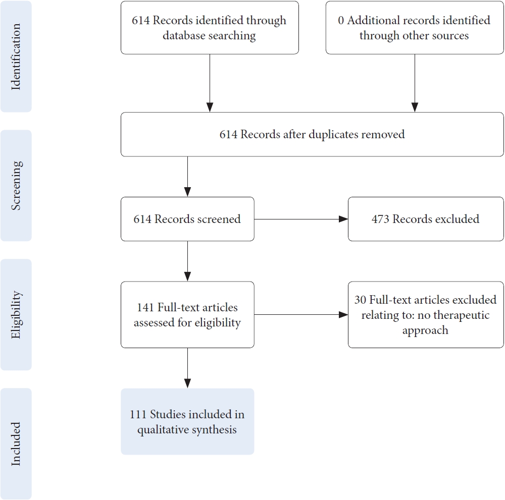
We performed a systematic, qualitative review of the literature, by screening the established MEDLINE databases PubMed, Embase, and OVID MEDLINE. The main search terms included “spinal cord injury,” angiogenesis,” “angiogenesis inducing agents,” and “tissue engineering,” in combination with “rodent subjects.” The search included no time limit. Articles in English were included, meeting the following criteria: experimental research, original studies, rodent subjects, therapeutic intervention, and focus on vascular injury and repair. The systematic literature search was conducted by one researcher from June 2021 to July 2021 and from May 2022 to June 2022. Data analysis was conducted according to the PRISMA (Preferred Reporting Items for Systematic Reviews and Meta-Analyses) guidelines. A total of 614 articles were selected for review, of which 475 were excluded after screening as a result of not meeting the inclusion criteria. An additional 30 articles were excluded due to not meeting the inclusion criteria after full-text assessment for eligibility. For qualitative synthesis 111 articles were assessed in detail and included in this publication (Fig. 1).

2. Relevant Experimental Therapeutic Approaches According to the Literature Search

According to the systematic literature search, 5 main categories of therapeutic approaches to promote vascular repair in experimental SCI research were identified: (1) angiogenic factors, (2) genetic engineering, (3) physical stimulation, (4) cell transplantation, and (5) biomaterials carrying various factor delivery (Table 1).

RESULTS

1. PART 1: Pathophysiology of Traumatic SCI With Emphasis on Vascular Injury And Repair

1) Pathophysiology and its progression following SCI

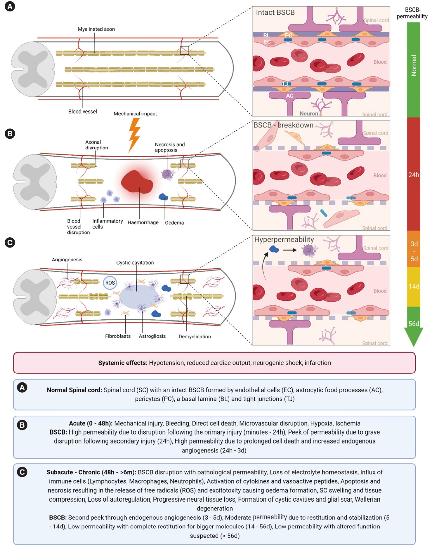
SCI is characterized as a sudden mechanical force impacting the spinal cord, e.g., following a fall, vehicle accident, or sportsrelated injury. It is commonly comprised of both contusion and compression injury causing direct cell death, axonal shearing, and disruption of the microvasculature with traumatic bleeding. This primary injury is not accessible to treatment [1,12] and is immediately followed by a sustained secondary injury cascade [13]. This secondary injury spreads to formerly unaffected regions, leading to further spinal cord damage with the consequences of demyelination and axonal dieback [2,3,14]. The developing secondary injury can be divided into acute (first 48 hours), subacute (48 hours–14 days), intermediate (14 days to 6 months) and chronic phases (> 6 months) [2,12] (Fig. 2).

2) Time course of BSCB disruption and restitution following SCI

The BSCB protects the spinal cord, as part of the central nervous system (CNS), from the periphery and maintains a homeostatic environment. The BSCB, formed by endothelial cells (ECs), astrocytic foot processes and pericytes as well as a basal lamina together with adjoining neurons comprise the neurovascular unit (NVU) (Fig. 2A). The unique cell-cell-contacts between ECs containing CNS-specific tight junctions (TJs) provide an immune privilege to the spinal cord compared to the periphery, restricting the passage of neurotoxic molecules and inflammatory cells [2,15].
Following the primary injury, a localized direct disruption of the NVU damages the microvasculature directly surrounding the injury site: Disruption of the protective cell-cell-contacts leads to altered cell permeabilization and consequent oedema formation, causing increased apoptotic and necrotic cell death [14,16,17] (Fig. 2B). These permeability changes not only lead to electrolyte influx and oedema, but to an influx of inflammatory cells (lymphocytes, macrophages, and neutrophils), as well as cytokines (tumor necrosis factor-α, interleukin [IL]-1β, IL-5) and vasoactive peptides. This immune response, commencing within the first minutes postinjury, leads to further oedema formation and thus tissue compression, resulting in further injury development spreading farther from the primary injury site [2,15,18]. These events further destabilize the BSCB and lead to a release of reactive oxygen species, resulting in the dysfunction of the NVU in formerly unharmed regions [2,3,14]. Correlating with these mechanisms, the first peak in BSCB-permeability in the penumbra zone is reached 24-hour postinjury, followed by a smaller peak after 3–5 days [4,5].
Most of the BSCB function is believed to be reinstated after 14 days [4,19] (Fig. 2C). However, experimental studies show that the permeability for small molecules is increased up to the chronic phase of SCI, for as long as 56-day postinjury [6].

3) Endogenous revascularization after SCI

The major form of vascular regeneration in the spinal cord is known to be angiogenesis, with a smaller part displayed by postnatal vasculogenesis. Angiogenesis is defined by the formation of blood vessels out of pre-existing vessels, whilst vasculogenesis describes the formation of new vessels [20]. The hypoxic milieu after SCI results in the release of angiogenic factors from the injury site. Endogenous revascularization following SCI is expected to occur during the first week postinjury, and to disappear shortly afterwards [21-24]. Importantly, the second, smaller peak in BSCB-permeability correlates with the peak of endogenous revascularization after SCI. As new vessels form due to angiogenesis or vasculogenesis, this young vasculature does not necessarily possess a functional BSCB. As some parts of this newly formed vasculature gain a functional barrier and promote a functional NVU during its genesis, other parts never do [4,15]. This results in the increased extravasation of molecules and fluids causing oedematous swelling, inflammation, and necrotic and apoptotic cell death and thus exacerbating the secondary injury [2,3,5,25] (Fig. 2B).

4) Regenerative potential facilitated by vascular repair after SCI

As the spinal cord remains in a state of chronic hypoxia and ischemia postinjury, reinstatement of a steady tissue perfusion would be desirable. This was shown by previous research indicating that neovascularization is crucial for axon regeneration, as these were shown to grow along blood vessels. This suggests that the developing vasculature guides axonal sprouting after injury [7,8]. However, the increased permeability for molecules otherwise excluded from the selective CNS environment might provide a unique chance for therapeutic intervention, with a potential window of opportunity displayed over the first days postinjury [4,26].
To address this multifaceted pathophysiology, several aspects of SCI must be taken into account to allow for tissue regeneration: Inhibition of the inflammatory response and sufficient perfusion might limit the secondary injury, while prevention of the glial scar formation could enable neural regeneration. Therefore, combinatorial approaches accounting for these mechanisms together in the adequate timespan might be promising for spinal cord repair.

2. PART 2: Experimental Therapeutic Approaches To Promote Vascular Repair

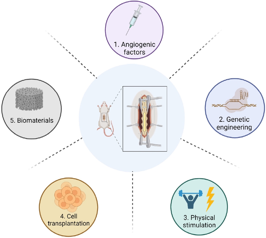
According to the systematic literature search, several experimental therapeutic approaches promising to diminish hypoxia and improve spinal cord microvasculature after SCI exist. In summary, 5 categories of experimental therapy were identified: (1) delivery of angiogenic factors, (2) genetic engineering, (3) physical stimulation, (4) cell transplantation, and (5) biomaterials carrying various factor delivery [9] (Fig. 3). In the following section of this study, we aim to provide a comprehensive overview of the literature in this field, comparing and discussing the most promising approaches for clinical translation.

1) Angiogenic factors

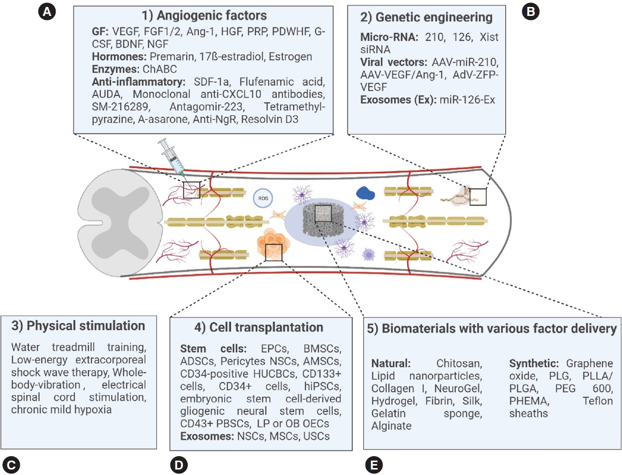
Many different therapeutic agents evoking an angiogenic answer, either directly or indirectly, have been investigated in experimental SCI models, often in combination with the implantation of biomaterials. In the following, we give an overview of different angiogenic factors and their previous experimental use in SCI research (Fig. 4A).
The therapeutic effects of vascular endothelial growth factor (VEGF) in SCI have been variable. VEGF-A is used in many studies and is a promising growth factor that binds to the tyrosine kinase receptors (VEGFRs) 1 and 2, resulting in the proliferation of ECs and angiogenesis. VEGF-A may also have neurotrophic, neuroprotective, and neuroproliferative effects [27]. A common application of VEGF-A in experimental SCI is the direct injection into the lesion site which results in increased blood vessel density and blood vessel diameter [28-29]. This effect has been confirmed in studies of controlled release of VEGF from biomaterials, such as gel foam [30]. On the downside, increased BSCB-permeability was observed after application of VEGF-A indicating the growth of non- or only partially functional blood vessels. Changes in vessel architecture, resulting in tumor-like blood vessels, were described in human studies [27,31].
To improve the angiogenic and neurotrophic effect through the application of VEGF, a release from biomaterials in combination with other growth factors such as fibroblast growth factor-2 (FGF2) [32], angiopoietin 1 and FGF [2,33] or brain-derived neurotrophic factor [34] was explored, also resulting in increased blood vessel density and associated localization of mature oligodendrocytes and axons.
FGF2 has also been frequently applied in experimental SCI. FGF2 was found to be highly overexpressed after SCI [35] and FGF2 is known to play a role in proliferation, differentiation and BSCB-integrity [36,37]. The application of FGF2 via biomaterials has been shown to increase the number of blood vessels [38-40], whereas no significant vascular changes were observed when FGF2 was infused at the injury site [41].
Further angiogenic factors that were previously tested for application in experimental SCI are hepatocyte growth factor (HGF) and the whole blood concentrates platelet-rich plasma (PRP) and platelet-derived wound healing formula (PDWHF). The number of blood vessels has been shown to increase after the application of activated PRP [42], PDWHF alone, or PDWHF in combination with nerve growth factor and of HGF [42-44].
Apart from these often-applied factors, other factors such as hormones, especially oestrogen [45], enzymes [46], and a multitude of anti-inflammatory substances [47] listed in (Fig. 4A) also showed potential to induce angiogenesis or to increase BSCB-integrity following SCI.

2) Genetic engineering

Genetic engineering is not widely used in experimental SCI but was shown to hold the potential to increase vascular repair and functional outcome after SCI [26,48-51] (Fig. 4B). Proofs-of-concept were brought forward in experiments using RNAs like micro-RNA-210 [48], micro-RNA-126 contained in exosomes [49] and X-inactive specific transcript-RNA [50]. The endogenous overexpression of VEGF using viral vectors also increased the number of blood vessels at the injury site [26,51]. The combination of VEGF165 and Ang-1 using an adeno-associated viral vector furthermore decreased the BSCB-permeability at the injury site [51]. Therefore, genetic engineering could become a promising approach to support vascular repair after experimental SCI [51].

3) Physical stimulation

As the only noninvasive therapeutic approach, physical stimulation was also shown to increase angiogenesis and functional outcome after experimental SCI. Methods under investigation include water treadmill training [52], low-energy extracorporeal shock wave therapy [53], whole-body vibration [54], chronic mild hypoxia [55], and electric stimulation [56] (Fig. 4C). Especially water treadmill training seems to improve the regeneration of functional blood vessels and improve functional outcome after SCI [52]. Using low-energy extracorporeal shock wave therapy, not only increased vessel density but also enhanced functional outcomes suggesting that the regenerative potential is higher than the risk of further damage to the spinal cord. Electrical stimulation has made tremendous progress as experimental therapy in both preclinical and clinical studies, although only few studies focus on the evaluation of angiogenic effects. The application of transspinal direct current stimulation increases the blood temporarily and is able to stimulate cell proliferation and migration [56]. Another experimental therapeutic approach is the induction of chronic mild hypoxia after SCI. The induction of mild hypoxia results in vascular remodelling, with endothelial proliferation and vascular expansion being more pronounced in the white matter. Interestingly, newly formed blood vessels grow towards neurons and show the upregulation of TJ proteins, indicating the enhanced formation of functional blood vessels [55]. These results are promising to increase angiogenic repair after SCI, but further research needs to address the effect of these approaches in the intermediate and chronic injury phase.

4) Cell transplantation

Cell transplantation is widely used after experimental SCI. Stem cells of different origins can result in a significant increase in angiogenesis and have previously been injected, infused, or implanted following SCI with the result of potently increasing the angiogenic response and improving functional outcome (Fig. 4D). Endothelial progenitor cells (EPCs) [57], bone marrow mesenchymal stem cells [58], adipose-derived stem cells [59], human umbilical cord blood stem cells [60], neural stem cells (NSCs) [61], amniotic mesenchymal stem cells [62,63], human-induced pluripotent stem cells [64,65], peripheral blood stem cells [66], lamina propria-derived olfactory ensheathing cells, olfactory bulb-derived ensheathing cells [67], and mesenchymal stem cells (MSCs) [68] increase angiogenesis when applied after SCI by expressing a wide range of growth factors and modulating the microenvironment. The effect of these cell types is shown in Table 2.
As not all implanted cells survive the hypoxic environment at and around the injury site, cell-based therapies to date can only unfold a fraction of their potential [69]. To overcome this limitation, other strategies rely on indirectly utilizing the therapeutic potential of stem cells. Especially the isolation of exosomes from different stem cells and their injection after experimental SCI showed similar results in terms of angiogenesis and functional outcome [70-72]. Exosomes derived from many different cell types result in increased angiogenesis after injection. Amongst these are NSC-derived exosomes [70], exosomes derived from human placenta-derived MSCs [71], and mesenchymal stromal cells-derived exosomes [72].
Another promising strategy is the combinatorial release of stem cells via biomaterials to enhance cell survival at the injury site by modulating the environment and by providing guidance for the regenerating tissue [69,73].

5) Biomaterials carrying various factor delivery

Biomaterials are materials engineered to interact with biological tissue and can be divided into natural materials sourced from plants or animals, synthetic materials and hybrid biomaterial combinations [10]. Many different biomaterials improve angiogenesis when implanted in combination with stem cells or angiogenic factors or even without concomitant application of other therapeutics. The implantation of biomaterials alone already holds the potential to support the ingrowth of blood vessels into the lesion site. This angiogenic potential was shown using many different biomaterials, such as a reduced graphene oxide scaffold [74], an oxygen-generating hydrogel scaffold [75,76], a nanofiber-hydrogel composite [77], “NeuroGel” [78], an aligned fibrin hydrogel [79], and a collagen type I scaffold [80] (Fig. 4E). Biomaterials can be combined with other therapeutics at will, such as angiogenic factors and/or stem cells to potentiate their regenerative potential.
Both natural and synthetic biomaterials were previously used successfully in animal studies to promote vascular repair after SCI with different advantages and disadvantages. Natural biomaterials are often intrinsically biodegradable and therefore useful to modify the delayed release of angiogenic factors or cells [81]. Synthetic biomaterials, unless modified accordingly, are nondegradable and guarantee more structural and long-lasting stability as well as reduced batch-to-batch variance. For implantation and combination with factor delivery, they can be sterilized and chemically modified to allow for an optimal release profile [10,81] (Table 3). Due to their high modifiability, both natural and synthetic biomaterials can be adapted to provide optimal release kinetics of incorporated factors at a defined location [10]. Release kinetics can be modified by using the intrinsic degradation rate of the biomaterial itself [82] or noncovalent interactions between the incorporated therapeutics and the biomaterial [40].
After implantation or injection, biomaterials can occupy the space of a posttraumatic cavity and provide a cell-friendly environment with reduced infiltration of inflammatory cells [77,81]. Therefore, biomaterials are often used in combination with stem cells, which highly depend on such an environment [83].
Exemplary, the number and density of blood vessels could be increased after implantation of an oligopolyethylene-glycol-fumarate-hydrogel combined with Rapamycin and Schwann-cells [83], of a prevascularized poly-L-lactic acid (PLLA)–polylactide-co-glycolide acid (PLGA) scaffold containing dental pulp stem cells [84], a fibrous porous silk scaffold containing human umbilical vein ECs [85], a hydrogel PLGA-scaffold containing NSCs and ECs [73], a gelatine sponge containing MSCs [69], and a PLGA-scaffold containing human MSCs [86]. The implantation of a hydrogel containing NSCs and EPCs also increased the formation of blood vessels inside the scaffold [87]. These findings show the great potential of combinatorial approaches using biomaterials containing angiogenic factors and/or stem cells of different types.
The application of biomaterials to promote vascular repair after SCI varies in time, location, and mode of application in the analysed studies (Table 3, Supplementary Table 1). In nearly all studies showing vascular changes, biomaterials are applied directly after induction of the injury with monitoring for up to 8 months [30,32-34,38-40,43,44,47,57,69,73-76,78,80,82-104]. Only one study reports an increased blood vessel density after the implantation of a nanofiber-hydrogel composite 3 days after injury [77]. Biomaterials are mostly applied at the injury site as scaffolds replacing the lesion after implantation [32,34,38,40,43,44,47,69,73,74,78,80,83-90,92-94,96-105] or injection[33,75,77,82,95] with only a few hydrogels being implanted on top of the injured spinal cord [30,39,91] or injected systemically [76].

POTENTIAL FOR CLINICAL IMPLEMENTATION

Taken together, all 5 experimental approaches discussed in this review show the potential to improve vascular repair and angiogenesis. The application of angiogenic factors (1) is a well-studied approach. Especially growth factors like VEGF have shown a promising potential to regenerate functional blood vessels. Genetic engineering (2) allows for direct activation of the endogenous expression of VEGF and other growth factors. Even if it is not applied as frequently as other approaches after SCI, it holds the potential to regenerate functional blood vessels. As physical stimulation techniques (3) also displayed the ability to induce vascular repair, they might be a useful supplementary therapy in a clinical setting as soon as intensive rehabilitation is possible. Cell transplantation (4) is another promising strategy to regenerate functional blood vessels after traumatic SCI, especially as they can be taken from a variety of sources. Their potential is inhibited by the toxic environment, which needs to be overcome to allow the cells to integrate at the injury site. The implantation of cells still poses a promising approach when combined with the implantation of biomaterials (5). These materials can be taken from many sources and are easily modifiable. Some have an inherent angiogenic potential and are biodegradable. As they can be modified to release incorporated therapeutics in the desired time frame at defined locations and provide guidance for regenerating axons, these combinatorial approaches seem promising for future research and potential clinical implementation. But before implementing promising biomaterials into clinical trials, several questions need to be addressed. For most biomaterials, especially when integrated in a combinatorial approach, the exact degradation time in SCI is neither known for humans nor for animals. As degradation byproducts might cause immunological reactions, biomaterials need to be actively characterized in preclinical and clinical studies [106]. Furthermore, most of the biomaterials summarized are solid materials filling a transection or hemisection injury, but contusion SCI is most common clinically. This and the overall limited comparability from controlled animal models into the individual human SCI makes it difficult to translate the results of preclinical studies into the design of clinical trials and shows the need for using clinically relevant SCI animal models with contusion or compression injuries.
Before applying these approaches in a clinical setting, future research needs to evaluate the ideal combinations to accurately address the multifaceted pathophysiology and the time course of BSCB de- and regeneration to allow for the regeneration of functional blood vessels as a basis for neural regeneration.

CONCLUSION

SCI is characterized by the injury of both neural and vascular components and results in extensive tissue loss. The disruption of the vasculature and its insufficient regeneration with a prolonged state of BSCB-permeability exacerbate the primary injury by amplification of secondary injury mechanisms. To restore a functional vasculature to prevent neural loss and to promote neural regeneration is one of the most important challenges in SCI research. Several therapeutic approaches show an improved vascularization after SCI, like the application of angiogenic factors, genetic engineering, physical stimulation, cell transplantation, and biomaterials carrying various factor delivery. Combinatorial approaches, like implanted biomaterials with the ability to release angiogenic factors or therapeutic cells in a temporally and spatially controlled manner seem most promising to restore functional vasculature and to be translatable into clinical patient care in the future.

SUPPLEMENTARY MATERIALS

Supplementary Table 1 can be found via https://doi.org/10.14245/ns.2244624.312.
Supplementary Table 1.
Complete list of studies using biomaterials to promote vascular repair after spinal cord injury identified by the systematic literature search.
ns-2244624-312-suppl.pdf

NOTES

Conflict of Interest

The authors have nothing to disclose.

Funding/Support

This study received no specific grant from any funding agency in the public, commercial, or not-for-profit sectors.

Author Contribution

Conceptualization: LR, VH, MGF, PV; Data curation: LR, VH; Formal analysis: LR, VH; Project administration: VH, MGF; Visualization: LR; Writing - original draft: LR, VH; Writing - review & editing: VH, JS, LKS, MGF

ACKNOWLEDGEMENTS

Figures were created using BioRender.com.

Fig. 1.
Flow chart of the conducted literature review and the systematic synthesis according to the PRISMA (Preferred Reporting Items for Systematic Reviews and Meta-Analyses) guidelines for systematic reviews and meta-analyses.
ns-2244624-312f1.jpg
Fig. 2.
(A-C) Pathophysiology of spinal cord injury and the neurovascular unit in health and its disruption. SC, spinal cord; BSCB, blood – spinal cord barrier; EC, endothelial cells; AC, astrocytic foot processes; PC, pericyte; BL, basal lamina; TJ, tight junction; ROS, reactive oxygen species; h, hours; d, days; w, weeks; m, months.
ns-2244624-312f2.jpg
Fig. 3.
Therapeutic approaches for vascular repair after experimental spinal cord injury: (1) angiogenic factors, (2) genetic engineering, (3) physical stimulation, (4) cell transplantation, and (5) biomaterials carrying various factor delivery.
ns-2244624-312f3.jpg
Fig. 4.
(A-E) Overview of therapeutic approaches inducing spinal cord repair after experimental spinal cord injury. PLG(A), polylactide-co-glycolide (acid); PLLA, poly-l-lactic acid; PEG600, polyethylene glycol 600; PHEMA, poly(2-hydroxyethyl methacrylate); VEGF, vascular endothelial growth factor; FGF, fibroblast growth factor; Ang-1, angiopoietin-1; HGF, hepatocyte growth factor; PRP, platelet-rich plasma; PDWHF, platelet-derived wound healing formula; G-CSF, granulocyte colony-stimulating factor; BDNF, brain-derived neurotrophic factor; NGF, nerve growth factor; ChABC, chondroitinase ABC; SDF-1a, stromal cellderived factor-1 alpha; AUDA, soluble epoxide hydrolase inhibitor; Anti-NgR, anti-nogo receptor antibody; Xist, X-inactive specific transcript; AAV, adeno-associated virus; AdV-ZFP-VEGF, adenoviral vector with zinc-finger protein coding for VEGF; EPC, endothelial progenitor cell; BMSC, bone mesenchymal stem cell; ADSC, adipose-derived stem cell; HUCBC, human umbilical cord blood stem cell; NSC, neural stem cell; AMSC, amniotic mesenchymal stem cell; hiPSC, human-induced pluripotent stem cell; PBSC, peripheral blood stem cell; LP OEC, lamina propria-derived olfactory ensheathing cell; OB OEC, olfactory bulbderived ensheathing cell; MSC, mesenchymal stem cell; USC, human urine stem cell.
ns-2244624-312f4.jpg
Table 1.
Therapeutic approaches for vascular repair after experimental spinal cord injury
Therapeutic approaches No. of included studies
1. Angiogenic factors 25
2. Genetic engineering 8
3. Physical stimulation 8
4. Cell transplantation 28
5. Biomaterial implantation 42
Table 2.
Study overview of experimentally transplanted cells to promote vascular repair after experimental spinal cord injury (SCI)
Species SCI model Method Outcome References
Rat T10 Contusion EPCs Increased axonal and blood vessel regeneration with functional improvement. Wang et al. [57] (2018)
Rat T10 Contusion BMSCs overexpressing VEGF Increased axonal and blood vessel regeneration with functional improvement. Liu et al. [58] (2020)
Increased VEGF protein expression.
Rat T8, 9 Compression ADSCs Increased blood vessel regeneration, vascular branching, and axonal regeneration. Menezes et al. [107] (2020)
Increased formation of functional blood vessels, which are associated with blood vessels.
Functional improvement.
Rat T10 Contusion HUCBCs Increased blood vessel regeneration with functional improvement. Ning et al. [60] (2013)
Rat T8 – 10 Contusion NSCs Increased blood vessel regeneration, VEGF protein expression, and remyelination with functional improvement. Li et al. [61] (2014)
Rat T9 Contusion AMSCs Increased blood vessel regeneration, axonal regeneration, VEGF protein expression, and functional improvement. Zhou et al. [62,63] (2016, 2020)
The BSCB – disruption was reduced at 7 days indicating the regeneration of functional vessels.
Rat T10 Contusion hiPSCs Increased blood vessel regeneration, myelination, and axonal regeneration with functional improvement. Transplanted cells differentiated into astrocytes, oligodendrocytes, and neurons which integrated with the host neural circuitry. Nori et al. [64] (2011)
Mice T9 Contusion PBSCs Increased blood vessel regeneration, myelination, and axonal regeneration with functional improvement. Takahashi et al. [66] (2016)
Rat C3, 4 Dorsolateral crush LP and OB OECs Increased axonal and blood vessel regeneration. Richter et al. [67] (2005)
The motor function was not assessed.
Rat T8 Contusion MSCs Increased axonal and blood vessel regeneration with no functional improvement. Kumagai et al. [68] (2013)

EPC, endothelial progenitor cell; BMSC, bone mesenchymal stem cell; VEGF, vascular endothelial growth factor; ADSC, adipose-derived stem cell; HUCBC, human umbilical cord blood stem cell; NSC, neural stem cell; AMSC, amniotic mesenchymal stem cell; hiPSC, human-induced pluripotent stem cell; PBSC, peripheral blood stem cell; LP OEC, lamina propria-derived olfactory ensheathing cell; OB OEC, olfactory bulbderived ensheathing cell; MSC, mesenchymal stem cell.

Table 3.
Study overview of experimentally used biomaterials to promote vascular repair after experimental spinal cord injury (SCI)
Species SCI model Method Outcome References
Natural biomaterials
Rat Lateral hemisection NSCs, EPCs, hydrogel NSCs and blood vessels infiltrate the hydrogel functional improvement Marrotte et al. [87] (2021)
Rat Lateral hemisection AFG/fSAP hydrogel Increased axonal regeneration with more myelinated axons, blood vessel regeneration at the lesion site with a larger diameter, increased vessel density, blood vessels and axons colocalized, functional improvement Man et al. [101] (2021)
Rat Compression injury CORM-2-lipid nanoparticles Increased formation of functional blood vessels and reduced BSCB-permeability at 1 day postinjury, functional improvement Joshi et al. [76] (2020)
Synthetic biomaterials
Rat Lateral hemisection rGO scaffold Regeneration of functional blood vessels growing inside the scaffold, association with regenerating axons López-Dolado et al. [74] (2016)
Rat Transection DPSCs PLLA/PLGA-scaffold Increase in axonal regeneration with myelin sheaths and angiogenesis at the injury site, vessel density increased in the rubrospinal tract, spinothalamic tract, dorsal column, and spinocerebellar tract, but not in corticospinal tract, functional improvement Guo et al. [84] (2020)
Rat Contusion VEGF, Ang-1, FGF2, PLGA-microspheres Increased angiogenesis and neural regeneration with axons and mature oligodendrocytes mostly associated with blood vessels, functional improvement Yu et al. [33] (2016)

NSC, neural stem cell; EPC, endothelial progenitor cell; AFG, aligned fibrin hydrogel; fSAP, functionalized self-assembling peptides derived from VEGF and brain-derived neurotrophic factor; CORM-2, carbon monoxide-releasing molecule-2; BSCB, blood-spinal cord barrier; rGO, reduced graphene oxide; DPSC, dental pulp stem cell; PLLA, poly-L-lactic acid; PLGA, polylactide-co-glycolide acid; VEGF, vascular endothelial growth factor; Ang-1, angiopoietin-1; FGF2, fibroblast growth factor 2.

REFERENCES

1. Ahuja CS, Nori S, Tetreault L, et al. Traumatic spinal cord injury-repair and regeneration. Neurosurgery 2017;80:S9-22.
crossref pmid pdf
2. Tator CH. Review of experimental spinal cord injury with emphasis on the local and systemic circulatory effects. Neurochirurgie 1991;37:291-302.
pmid
3. Tator CH. Update on the pathophysiology and pathology of acute spinal cord injury. Brain Pathol 1995;5:407-13.
crossref pmid
4. Figley SA, Khosravi R, Legasto JM, et al. Characterization of vascular disruption and blood-spinal cord barrier permeability following traumatic spinal cord injury. J Neurotrauma 2014;31:541-52.
crossref pmid pmc
5. Ng MT, Stammers AT, Kwon BK. Vascular disruption and the role of angiogenic proteins after spinal cord injury. Transl Stroke Res 2011;2:474-91.
crossref pmid pmc pdf
6. Sundberg LM, Herrera JJ, Narayana PA. In vivo longitudinal MRI and behavioral studies in experimental spinal cord injury. J Neurotrauma 2010;27:1753-67.
crossref pmid pmc
7. Bearden SE, Segal SS. Microvessels promote motor nerve survival and regeneration through local VEGF release following ectopic reattachment. Microcirculation 2004;11:633-44.
crossref pmid
8. Ohab JJ, Fleming S, Blesch A, et al. A neurovascular niche for neurogenesis after stroke. J Neurosci 2006;26:13007-16.
crossref pmid pmc
9. Yao C, Cao X, Yu B. Revascularization after traumatic spinal cord injury. Front Physiol 2021;12:631500.
crossref pmid pmc
10. Harvey AR, Lovett SJ, Majda BT, et al. Neurotrophic factors for spinal cord repair: which, where, how and when to apply, and for what period of time? Brain Res 2015;1619:36-71.
crossref pmid
11. Gadot R, Smith DN, Prablek M, et al. Established and emerging therapies in acute spinal cord injury. Neurospine 2022;19:283-96.
crossref pmid pmc pdf
12. Ahuja CS, Wilson JR, Nori S, et al. Traumatic spinal cord injury. Nat Rev Dis Primers 2017;3:17018.
crossref pmid pdf
13. Allen AR. Surgery of experimental lesion of spinal cord equivalent to crush injury of fracture dislocation of spinal column: a preliminary report. JAMA 1911;LVII:878-80.
crossref
14. Tator CH, Koyanagi I. Vascular mechanisms in the pathophysiology of human spinal cord injury. J Neurosurg 1997;86:483-92.
crossref pmid
15. Mautes AE, Weinzierl MR, Donovan F, et al. Vascular events after spinal cord injury: contribution to secondary pathogenesis. Phys Ther 2000;80:673-87.
crossref pmid pdf
16. Ling X, Bao F, Qian H, et al. The temporal and spatial profiles of cell loss following experimental spinal cord injury: effect of antioxidant therapy on cell death and functional recovery. BMC Neurosci 2013;14:146.
crossref pmid pmc pdf
17. Lou J, Lenke LG, Ludwig FJ, et al. Apoptosis as a mechanism of neuronal cell death following acute experimental spinal cord injury. Spinal Cord 1998;36:683-90.
crossref pmid pdf
18. Sandler AN, Tator CH. Review of the effect of spinal cord trama on the vessels and blood flow in the spinal cord. J Neurosurg 1976;45:638-46.
pmid
19. Noble LJ, Wrathall JR. Distribution and time course of protein extravasation in the rat spinal cord after contusive injury. Brain Res 1989;482:57-66.
crossref pmid
20. Risau W, Flamme I. Vasculogenesis. Annu Rev Cell Dev Biol 1995;11:73-91.
crossref pmid
21. Casella GT, Marcillo A, Bunge MB, et al. New vascular tissue rapidly replaces neural parenchyma and vessels destroyed by a contusion injury to the rat spinal cord. Exp Neurol 2002;173:63-76.
crossref pmid
22. Hagg T, Oudega M. Degenerative and spontaneous regenerative processes after spinal cord injury. J Neurotrauma 2006;23:264-80.
crossref pmid
23. Loy DN, Crawford CH, Darnall JB, et al. Temporal progression of angiogenesis and basal lamina deposition after contusive spinal cord injury in the adult rat. J Comp Neurol 2002;445:308-24.
crossref pmid
24. Ritz MF, Graumann U, Gutierrez B, et al. Traumatic spinal cord injury alters angiogenic factors and TGF-Beta1 that may affect vascular recovery. Curr Neurovasc Res 2010;7:301-10.
crossref pmid
25. Yip PK, Malaspina A. Spinal cord trauma and the molecular point of no return. Mol Neurodegener 2012;7:6.
crossref pmid pmc
26. Figley SA, Liu Y, Karadimas SK, et al. Delayed administration of a bio-engineered zinc-finger VEGF-A gene therapy is neuroprotective and attenuates allodynia following traumatic spinal cord injury. PLoS One 2014;9:e96137.
crossref pmid pmc
27. Greenberg DA, Jin K. From angiogenesis to neuropathology. Nature 2005;438:954-9.
crossref pmid pdf
28. Widenfalk J, Lipson A, Jubran M, et al. Vascular endothelial growth factor improves functional outcome and decreases secondary degeneration in experimental spinal cord contusion injury. Neuroscience 2003;120:951-60.
crossref pmid
29. van Neerven S, Joosten EA, Brook GA, et al. Repetitive intrathecal VEGF(165) treatment has limited therapeutic effects after spinal cord injury in the rat. J Neurotrauma 2010;27:1781-91.
crossref pmid
30. Patel CB, Cohen DM, Ahobila-Vajjula P, et al. Effect of VEGF treatment on the blood-spinal cord barrier permeability in experimental spinal cord injury: dynamic contrastenhanced magnetic resonance imaging. J Neurotrauma 2009;26:1005-16.
crossref pmid pmc
31. Benton RL, Whittemore SR. VEGF165 therapy exacerbates secondary damage following spinal cord injury. Neurochem Res 2003;28:1693-703.
pmid
32. De Laporte L, des Rieux A, Tuinstra HM, et al. Vascular endothelial growth factor and fibroblast growth factor 2 delivery from spinal cord bridges to enhance angiogenesis following injury. J Biomed Mater Res A 2011;98:372-82.
pmid pmc
33. Yu S, Yao S, Wen Y, et al. Angiogenic microspheres promote neural regeneration and motor function recovery after spinal cord injury in rats. Sci Rep 2016;6:33428.
crossref pmid pmc pdf
34. Wen Y, Yu S, Wu Y, et al. Spinal cord injury repair by implantation of structured hyaluronic acid scaffold with PLGA microspheres in the rat. Cell Tissue Res 2016;364:17-28.
crossref pmid pdf
35. Zai LJ, Yoo S, Wrathall JR. Increased growth factor expression and cell proliferation after contusive spinal cord injury. Brain Res 2005;1052:147-55.
crossref pmid
36. Zhu H, Yang A, Du J, et al. Basic fibroblast growth factor is a key factor that induces bone marrow mesenchymal stem cells towards cells with Schwann cell phenotype. Neurosci Lett 2014;559:82-7.
crossref pmid
37. Ye LB, Yu XC, Xia QH, et al. Regulation of caveolin-1 and junction proteins by bFGF contributes to the integrity of blood-spinal cord barrier and functional recovery. Neurotherapeutics 2016;13:844-58.
crossref pmid pmc pdf
38. Patist CM, Mulder MB, Gautier SE, et al. Freeze-dried poly(D,L-lactic acid) macroporous guidance scaffolds impregnated with brain-derived neurotrophic factor in the transected adult rat thoracic spinal cord. Biomaterials 2004;25:1569-82.
crossref pmid
39. Kang CE, Baumann MD, Tator CH, et al. Localized and sustained delivery of fibroblast growth factor-2 from a nanoparticle-hydrogel composite for treatment of spinal cord injury. Cells Tissues Organs 2013;197:55-63.
crossref pmid pdf
40. Shang J, Qiao H, Hao P, et al. bFGF-sodium hyaluronate collagen scaffolds enable the formation of nascent neural networks after adult spinal cord injury. J Biomed Nanotechnol 2019;15:703-16.
crossref pmid
41. Baffour R, Achanta K, Kaufman J, et al. Synergistic effect of basic fibroblast growth factor and methylprednisolone on neurological function after experimental spinal cord injury. J Neurosurg 1995;83:105-10.
crossref pmid
42. Chen NF, Sung CS, Wen ZH, et al. Therapeutic effect of platelet-rich plasma in rat spinal cord injuries. Front Neurosci 2018;12:252.
crossref pmid pmc
43. Hiraizumi Y, Fujimaki E, Transfeldt EE, et al. The effect of the platelet derived wound healing formula and the nerve growth factor on the experimentally injured spinal cord. Spinal Cord 1996;34:394-402.
crossref pmid pdf
44. Hiraizumi Y, Transfeldt EE, Kawahara N, et al. In vivo angiogenesis by platelet-derived wound-healing formula in injured spinal cord. Brain Res Bull 1993;30:353-7.
crossref pmid
45. Ni S, Cao Y, Jiang L, et al. Synchrotron radiation imaging reveals the role of estrogen in promoting angiogenesis after acute spinal cord injury in rats. Spine (Phila Pa 1976) 2018;43:1241-9.
crossref pmid
46. Milbreta U, von Boxberg Y, Mailly P, et al. Astrocytic and vascular remodeling in the injured adult rat spinal cord after chondroitinase ABC treatment. J Neurotrauma 2014;31:803-18.
crossref pmid pmc
47. Yao Y, Xu J, Yu T, et al. Flufenamic acid inhibits secondary hemorrhage and BSCB disruption after spinal cord injury. Theranostics 2018;8:4181-98.
crossref pmid pmc
48. Ujigo S, Kamei N, Hadoush H, et al. Administration of microRNA-210 promotes spinal cord regeneration in mice. Spine (Phila Pa 1976) 2014;39:1099-107.
crossref pmid
49. Huang JH, Xu Y, Yin XM, et al. Exosomes derived from miR-126-modified MSCs promote angiogenesis and neurogenesis and attenuate apoptosis after spinal cord injury in rats. Neuroscience 2020;424:133-45.
crossref pmid
50. Cheng X, Xu J, Yu Z, et al. LncRNA xist contributes to endogenous neurological repair after chronic compressive spinal cord injury by promoting angiogenesis through the miR-32-5p/notch-1 axis. Front Cell Dev Biol 2020;8:744.
crossref pmid pmc
51. Herrera JJ, Sundberg LM, Zentilin L, et al. Sustained expression of vascular endothelial growth factor and angiopoietin-1 improves blood-spinal cord barrier integrity and functional recovery after spinal cord injury. J Neurotrauma 2010;27:2067-76.
crossref pmid pmc
52. Shin HY, Kim H, Kwon MJ, et al. Molecular and cellular changes in the lumbar spinal cord following thoracic injury: regulation by treadmill locomotor training. PLoS One 2014;9:e88215.
crossref pmid pmc
53. Yahata K, Kanno H, Ozawa H, et al. Low-energy extracorporeal shock wave therapy for promotion of vascular endothelial growth factor expression and angiogenesis and improvement of locomotor and sensory functions after spinal cord injury. J Neurosurg Spine 2016;25:745-55.
crossref pmid
54. Manthou M, Nohroudi K, Moscarino S, et al. Functional recovery after experimental spinal cord compression and whole body vibration therapy requires a balanced revascularization of the injured site. Restor Neurol Neurosci 2015;33:233-49.
crossref pmid
55. Halder SK, Kant R, Milner R. Chronic mild hypoxia promotes profound vascular remodeling in spinal cord blood vessels, preferentially in white matter, via an α5β1 integrinmediated mechanism. Angiogenesis 2018;21:251-66.
crossref pmid pmc pdf
56. Samaddar S, Vazquez K, Ponkia D, et al. Transspinal direct current stimulation modulates migration and proliferation of adult newly born spinal cells in mice. J Appl Physiol (1985) 2017;122:339-53.
crossref pmid
57. Wang T, Fang X, Yin ZS. Endothelial progenitor cell-conditioned medium promotes angiogenesis and is neuroprotective after spinal cord injury. Neural Regen Res 2018;13:887-95.
crossref pmid pmc
58. Liu X, Xu W, Zhang Z, et al. Vascular endothelial growth factor-transfected bone marrow mesenchymal stem cells improve the recovery of motor and sensory functions of rats with spinal cord injury. Spine (Phila Pa 1976) 2020;45:E364-72.
crossref pmid
59. Oh JS, Park IS, Kim KN, et al. Transplantation of an adipose stem cell cluster in a spinal cord injury. Neuroreport 2012;23:277-82.
crossref pmid
60. Ning G, Tang L, Wu Q, et al. Human umbilical cord blood stem cells for spinal cord injury: early transplantation results in better local angiogenesis. Regen Med 2013;8:271-81.
crossref pmid
61. Li Z, Guo GH, Wang GS, et al. Influence of neural stem cell transplantation on angiogenesis in rats with spinal cord injury. Genet Mol Res 2014;13:6083-92.
crossref pmid
62. Zhou HL, Fang H, Luo HT, et al. Intravenous administration of human amniotic mesenchymal stem cells improves outcomes in rats with acute traumatic spinal cord injury. Neuroreport 2020;31:730-6.
crossref pmid
63. Zhou HL, Zhang XJ, Zhang MY, et al. Transplantation of human amniotic mesenchymal stem cells promotes functional recovery in a rat model of traumatic spinal cord injury. Neurochem Res 2016;41:2708-18.
crossref pmid pdf
64. Nori S, Okada Y, Yasuda A, et al. Grafted human-induced pluripotent stem-cell-derived neurospheres promote motor functional recovery after spinal cord injury in mice. Proc Natl Acad Sci U S A 2011;108:16825-30.
crossref pmid pmc
65. Kobayashi Y, Okada Y, Itakura G, et al. Pre-evaluated safe human iPSC-derived neural stem cells promote functional recovery after spinal cord injury in common marmoset without tumorigenicity. PLoS One 2012;7:e52787.
crossref pmid pmc
66. Takahashi H, Koda M, Hashimoto M, et al. Transplanted peripheral blood stem cells mobilized by granulocyte colony-stimulating factor promoted hindlimb functional recovery after spinal cord injury in mice. Cell Transplant 2016;25:283-92.
crossref pmid pdf
67. Richter MW, Fletcher PA, Liu J, et al. Lamina propria and olfactory bulb ensheathing cells exhibit differential integration and migration and promote differential axon sprouting in the lesioned spinal cord. J Neurosci 2005;25:10700-11.
crossref pmid pmc
68. Kumagai G, Tsoulfas P, Toh S, et al. Genetically modified mesenchymal stem cells (MSCs) promote axonal regeneration and prevent hypersensitivity after spinal cord injury. Exp Neurol 2013;248:369-80.
crossref pmid
69. Zeng X, Zeng YS, Ma YH, et al. Bone marrow mesenchymal stem cells in a three-dimensional gelatin sponge scaffold attenuate inflammation, promote angiogenesis, and reduce cavity formation in experimental spinal cord injury. Cell Transplant 2011;20:1881-99.
crossref pmid pdf
70. Zhong D, Cao Y, Li CJ, et al. Neural stem cell-derived exosomes facilitate spinal cord functional recovery after injury by promoting angiogenesis. Exp Biol Med (Maywood) 2020;245:54-65.
crossref pmid pmc pdf
71. Zhang C, Zhang C, Xu Y, et al. Exosomes derived from human placenta-derived mesenchymal stem cells improve neurologic function by promoting angiogenesis after spinal cord injury. Neurosci Lett 2020;739:135399.
crossref pmid
72. Huang JH, Yin XM, Xu Y, et al. Systemic administration of exosomes released from mesenchymal stromal cells attenuates apoptosis, inflammation, and promotes angiogenesis after spinal cord injury in rats. J Neurotrauma 2017;34:3388-96.
crossref pmid
73. Rauch MF, Hynes SR, Bertram J, et al. Engineering angiogenesis following spinal cord injury: a coculture of neural progenitor and endothelial cells in a degradable polymer implant leads to an increase in vessel density and formation of the blood-spinal cord barrier. Eur J Neurosci 2009;29:132-45.
crossref pmid pmc
74. López-Dolado E, Gonzalez-Mayorga A, Gutierrez MC, et al. Immunomodulatory and angiogenic responses induced by graphene oxide scaffolds in chronic spinal hemisected rats. Biomaterials 2016;99:72-81.
crossref pmid
75. Liu L, Wan J, Dai M, et al. Effects of oxygen generating scaffolds on cell survival and functional recovery following acute spinal cord injury in rats. J Mater Sci Mater Med 2020;31:115.
crossref pmid pdf
76. Joshi HP, Kumar H, Choi UY, et al. CORM-2-solid lipid nanoparticles maintain integrity of blood-spinal cord barrier after spinal cord injury in rats. Mol Neurobiol 2020;57:2671-89.
crossref pmid pdf
77. Li X, Zhang C, Haggerty AE, et al. The effect of a nanofiber-hydrogel composite on neural tissue repair and regeneration in the contused spinal cord. Biomaterials 2020;245:119978.
crossref pmid pmc
78. Woerly S, Doan VD, Sosa N, et al. Prevention of gliotic scar formation by NeuroGel allows partial endogenous repair of transected cat spinal cord. J Neurosci Res 2004;75:262-72.
crossref pmid pdf
79. Yao S, Yu S, Cao Z, et al. Hierarchically aligned fibrin nanofiber hydrogel accelerated axonal regrowth and locomotor function recovery in rat spinal cord injury. Int J Nanomedicine 2018;13:2883-95.
crossref pmid pmc pdf
80. Altinova H, Hammes S, Palm M, et al. Dense fibroadhesive scarring and poor blood vessel-maturation hamper the integration of implanted collagen scaffolds in an experimental model of spinal cord injury. Biomed Mater 2020;15:015012.
crossref pmid pdf
81. Haggerty AE, Maldonado-Lasuncion I, Oudega M. Biomaterials for revascularization and immunomodulation after spinal cord injury. Biomed Mater 2018;13:044105.
crossref pmid pdf
82. des Rieux A, De Berdt P, Ansorena E, et al. Vascular endothelial growth factor-loaded injectable hydrogel enhances plasticity in the injured spinal cord. J Biomed Mater Res A 2014;102:2345-55.
crossref pmid
83. Siddiqui AM, Oswald D, Papamichalopoulos S, et al. Defining spatial relationships between spinal cord axons and blood vessels in hydrogel scaffolds. Tissue Eng Part A 2021;27:648-64.
crossref pmid pmc
84. Guo S, Redenski I, Landau S, et al. Prevascularized scaffolds bearing human dental pulp stem cells for treating complete spinal cord injury. Adv Healthc Mater 2020;9:e2000974.
crossref pmid pdf
85. Zhong J, Xu J, Lu S, et al. A prevascularization strategy using novel fibrous porous silk scaffolds for tissue regeneration in mice with spinal cord injury. Stem Cells Dev 2020;29:615-24.
crossref pmid
86. Ropper AE, Thakor DK, Han I, et al. Defining recovery neurobiology of injured spinal cord by synthetic matrix-assisted hMSC implantation. Proc Natl Acad Sci U S A 2017;114:E820-9.
crossref pmid pmc
87. Marrotte EJ, Johnson K, Schweller RM, et al. Induction of neurogenesis and angiogenesis in a rat hemisection spinal cord injury model with combined neural stem cell, endothelial progenitor cell, and biomimetic hydrogel matrix therapy. Crit Care Explor 2021;3:e0436.
crossref pmid pmc
88. Brazda N, Estrada V, Voss C, et al. Experimental strategies to bridge large tissue gaps in the injured spinal cord after acute and chronic lesion. J Vis Exp 2016;(110):e53331.
crossref pmid
89. Liu D, Shen H, Shen Y, et al. Dual-cues laden scaffold facilitates neurovascular regeneration and motor functional recovery after complete spinal cord injury. Adv Healthc Mater 2021;10:e2100089.
crossref pmid pdf
90. Xi K, Gu Y, Tang J, et al. Microenvironment-responsive immunoregulatory electrospun fibers for promoting nerve function recovery. Nat Commun 2020;11:4504.
crossref pmid pmc pdf
91. Lutton C, Young YW, Williams R, et al. Combined VEGF and PDGF treatment reduces secondary degeneration after spinal cord injury. J Neurotrauma 2012;29:957-70.
crossref pmid
92. Xu ZX, Zhang LQ, Zhou YN, et al. Histological and functional outcomes in a rat model of hemisected spinal cord with sustained VEGF/NT-3 release from tissue-engineered grafts. Artif Cells Nanomed Biotechnol 2020;48:362-76.
crossref pmid
93. Bakshi A, Fisher O, Dagci T, et al. Mechanically engineered hydrogel scaffolds for axonal growth and angiogenesis after transplantation in spinal cord injury. J Neurosurg Spine 2004;1:322-9.
crossref pmid
94. Colello RJ, Chow WN, Bigbee JW, et al. The incorporation of growth factor and chondroitinase ABC into an electrospun scaffold to promote axon regrowth following spinal cord injury. J Tissue Eng Regen Med 2016;10:656-68.
crossref pmid
95. Gwak SJ, Yun Y, Yoon DH, et al. Therapeutic use of 3beta-[N-(N',N'-Dimethylaminoethane) Carbamoyl] cholesterol-modified PLGA nanospheres as gene delivery vehicles for spinal cord injury. PLoS One 2016;11:e0147389.
crossref pmid pmc
96. Wei YT, He Y, Xu CL, et al. Hyaluronic acid hydrogel modified with nogo-66 receptor antibody and poly-L-lysine to promote axon regrowth after spinal cord injury. J Biomed Mater Res B Appl Biomater 2010;95:110-7.
crossref pmid
97. Xu ZX, Zhang LQ, Wang CS, et al. Acellular spinal cord scaffold implantation promotes vascular remodeling with sustained delivery of VEGF in a rat spinal cord hemisection model. Curr Neurovasc Res 2017;14:274-89.
crossref pmid
98. Woerly S, Pinet E, de Robertis L, et al. Spinal cord repair with PHPMA hydrogel containing RGD peptides (NeuroGel). Biomaterials 2001;22:1095-111.
crossref pmid
99. Yang B, Wang PB, Mu N, et al. Graphene oxide-composited chitosan scaffold contributes to functional recovery of injured spinal cord in rats. Neural Regen Res 2021;16:1829-35.
crossref pmid pmc
100. Woerly S, Petrov P, Sykova E, et al. Neural tissue formation within porous hydrogels implanted in brain and spinal cord lesions: ultrastructural, immunohistochemical, and diffusion studies. Tissue Eng 1999;5:467-88.
crossref pmid
101. Man W, Yang S, Cao Z, et al. A multi-modal delivery strategy for spinal cord regeneration using a composite hydrogel presenting biophysical and biochemical cues synergistically. Biomaterials 2021;276:120971.
crossref pmid
102. Hakim JS, Rodysill BR, Chen BK, et al. Combinatorial tissue engineering partially restores function after spinal cord injury. J Tissue Eng Regen Med 2019;13:857-73.
crossref pmid pmc pdf
103. Li G, Che MT, Zeng X, et al. Neurotrophin-3 released from implant of tissue-engineered fibroin scaffolds inhibits inflammation, enhances nerve fiber regeneration, and improves motor function in canine spinal cord injury. J Biomed Mater Res A 2018;106:2158-70.
crossref pmid pmc pdf
104. Facchiano F, Fernandez E, Mancarella S, et al. Promotion of regeneration of corticospinal tract axons in rats with recombinant vascular endothelial growth factor alone and combined with adenovirus coding for this factor. J Neurosurg 2002;97:161-8.
crossref
105. Wang L, Shi Q, Dai J, et al. Increased vascularization promotes functional recovery in the transected spinal cord rats by implanted vascular endothelial growth factor-targeting collagen scaffold. J Orthop Res 2018;36:1024-34.
crossref pmid pdf
106. Jarrah R, Sammak SE, Onyedimma C, et al. The role of alginate hydrogels as a potential treatment modality for spinal cord injury: a comprehensive review of the literature. Neurospine 2022;19:272-80.
crossref pmid pmc pdf
107. Menezes K, Rosa BG, Freitas C, et al. Human mesenchymal stromal/stem cells recruit resident pericytes and induce blood vessels maturation to repair experimental spinal cord injury in rats. Sci Rep 2020;10:19604.
crossref pmid pmc pdf
  • GS_Medical
  • TOOLS
    Share :
    Facebook Twitter Linked In Google+
    METRICS Graph View
    • 10 Web of Science
    • 10 Crossref
    • 1 Scopus
    • 4,059 View
    • 181 Download
    Journal Impact Factor 3.8
    SURGERY: Q1
    CLINICAL NEUROLOGY: Q1
    Related articles in NS

    The Importance of Vascular Repair as the First Step in Spinal Cord Injury Treatment: Commentary on “Therapeutic Approaches Targeting Vascular Repair After Experimental Spinal Cord Injury: A Systematic Review of the Literature”2022 December;19(4)

    Commentary on “Effect of Acute Physical Interventions on Pathophysiology and Recovery After Spinal Cord Injury: A Comprehensive Review of the Literature”2022 September;19(3)

    Effect of Acute Physical Interventions on Pathophysiology and Recovery After Spinal Cord Injury: A Comprehensive Review of the Literature2022 September;19(3)

    The Role of Alginate Hydrogels as a Potential Treatment Modality for Spinal Cord Injury: A Comprehensive Review of the Literature2022 June;19(2)

    Curcumin as a Promising Neuroprotective Agent for the Treatment of Spinal Cord Injury: A Review of the Literature2022 June;19(2)


    Editorial Office
    CHA University, CHA School of Medicine Bundang Medical Center
    59 Yatap-ro, Bundang-gu, Seongnam-si, Gyeonggi-do 13496, Korea
    Tel: +82-31-780-1924  Fax: +82-31-780-5269  E-mail: support@e-neurospine.org
    The Korean Spinal Neurosurgery Society
    #407, Dong-A Villate 2nd Town, 350 Seocho-daero, Seocho-gu, Seoul 06631, Korea
    Tel: +82-2-585-5455  Fax: +82-2-2-523-6812  E-mail: ksns1987@gmail.com
    Business License No.: 209-82-62443

    Copyright © The Korean Spinal Neurosurgery Society.

    Developed in M2PI

    Zoom in Close layer